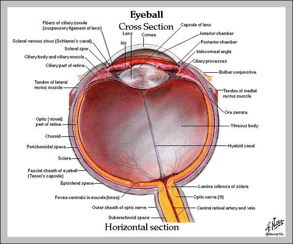
Labelled Diagram Of The Eye
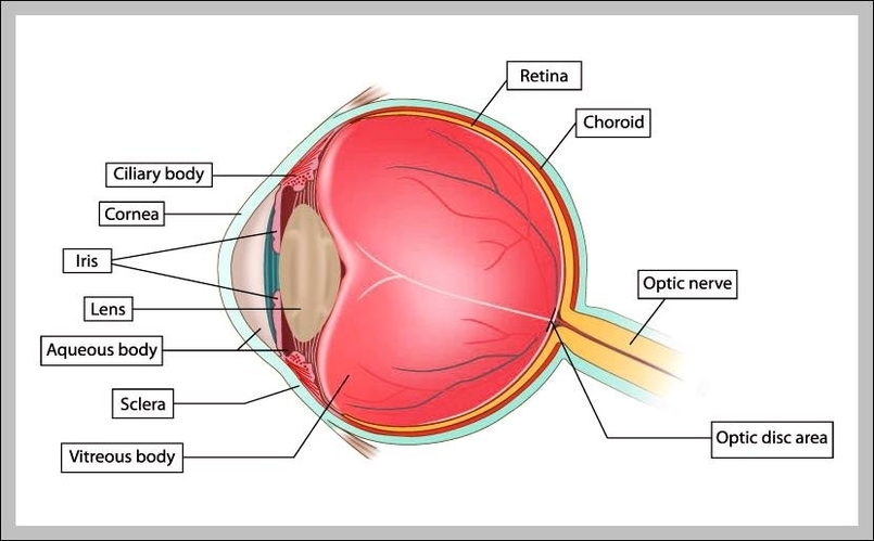
Labelled Diagram Of The Eye: A labeled eye diagram shows key structures such as the cornea, iris, pupil, lens, retina, and optic nerve, each contributing to the sense of sight.