
Knee Muscle

Knee Muscle: Knee muscles, primarily the quadriceps and hamstrings, work together to extend and flex the knee joint, playing a major role in walking, running, and stability.
Knee Muscle: Knee muscles, primarily the quadriceps and hamstrings, work together to extend and flex the knee joint, playing a major role in walking, running, and stability.
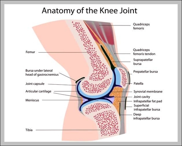
Knee Joint Capsule: The knee joint capsule is a fibrous tissue enclosing the knee joint, containing synovial fluid that lubricates and protects the joint during movement.

Knee Anatomy Muscles: The muscles around the knee include the quadriceps, hamstrings, and calf muscles, which all help with the flexion and extension of the knee joint.
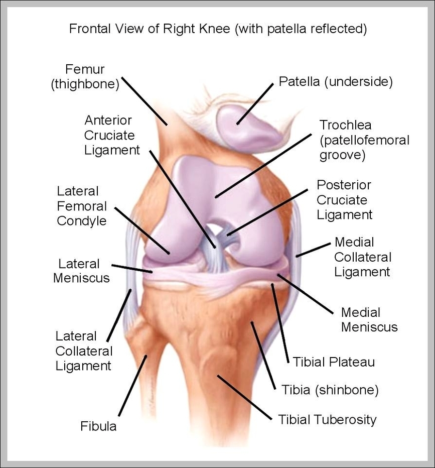
Knee Anatomy Diagram: A knee anatomy diagram shows bones (femur, tibia, patella), ligaments (ACL, PCL), tendons, and cartilage that allow the knee to function as a hinge joint, supporting mobility and weight.
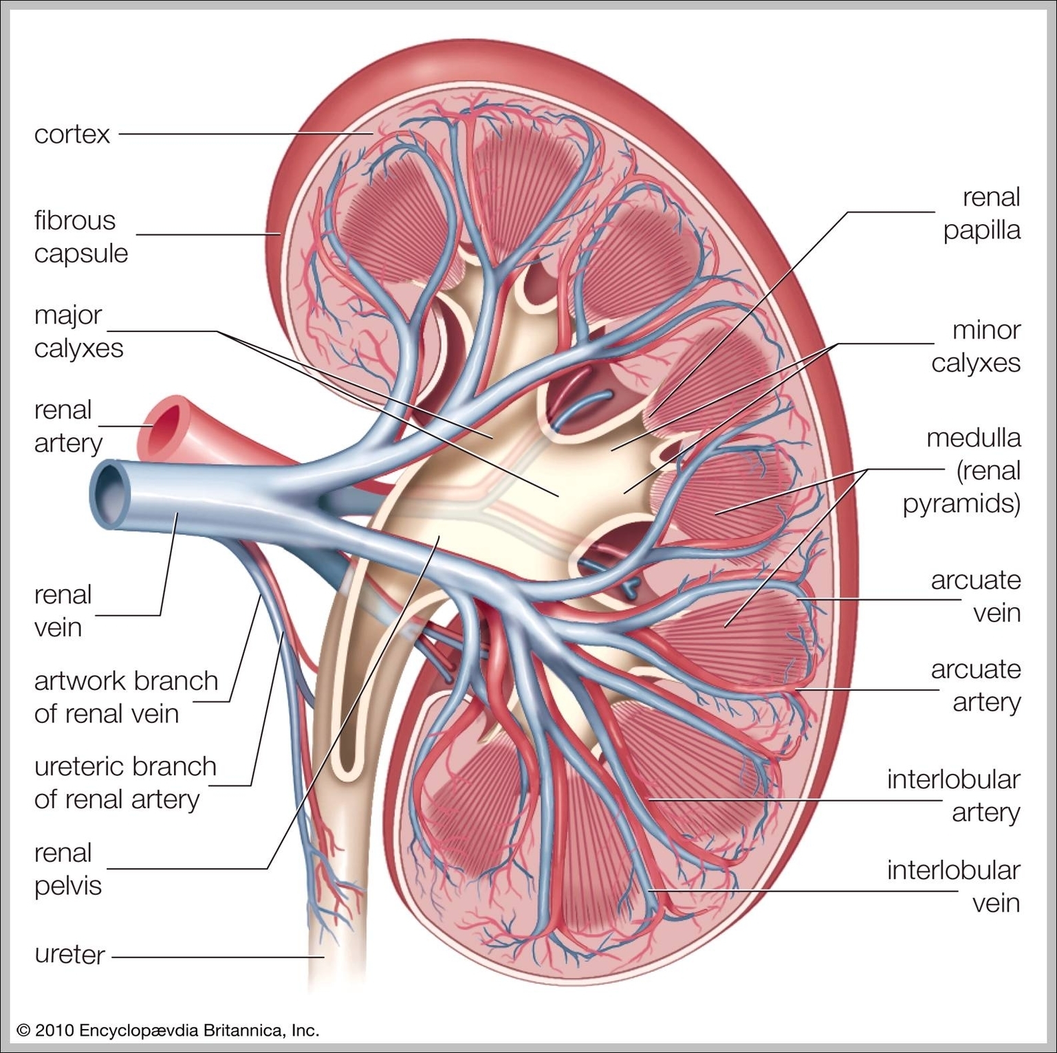
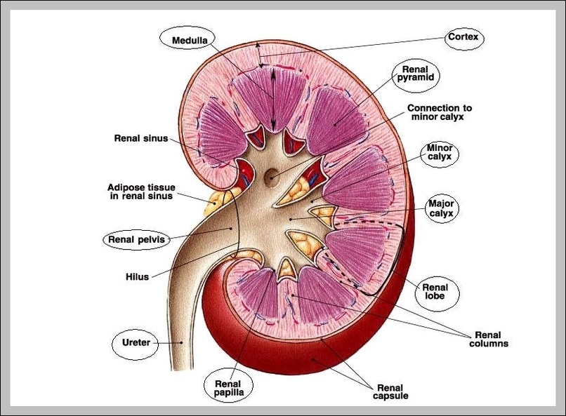
Kidney Tissue: Kidney tissue includes various layers such as the renal cortex and renal medulla, with specialized structures like nephrons that filter blood, removing waste and regulating fluid balance.
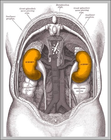
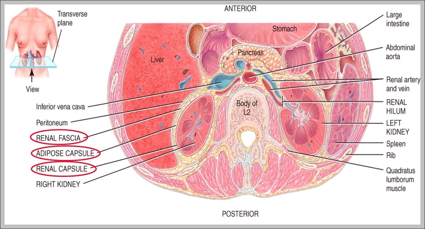
Kidneys Location In Human Body: The kidneys are located in the upper abdominal area, just below the ribcage on either side of the spine, behind the abdominal organs.
Kidneys Function: The kidneys are responsible for filtering blood, removing waste, balancing electrolytes, and regulating fluid levels in the body. They also produce hormones that regulate blood pressure and red blood cell production.
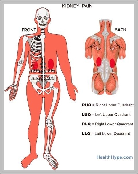
Kidney Location On The Body: The kidneys are located on either side of the spine, just below the ribcage. They are responsible for filtering blood, removing waste, and regulating fluid and electrolyte balance.
Kidney In Body Anatomy: In body anatomy, the kidneys are bean-shaped organs located on either side of the spine in the lower back, vital for filtering waste and balancing fluids.
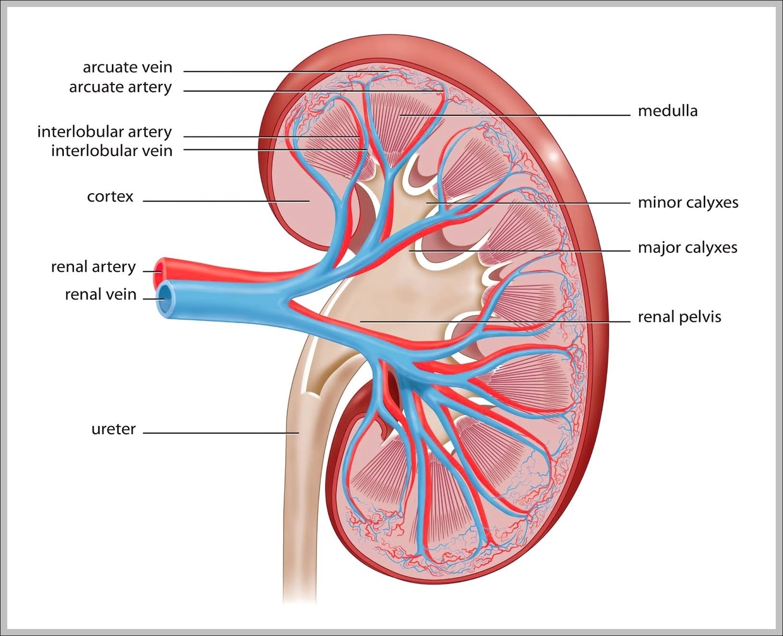
Kidney Image: A kidney image typically shows its bean shape, outer cortex, inner medulla, renal pelvis, and ureter, detailing its role in filtering blood and forming urine.
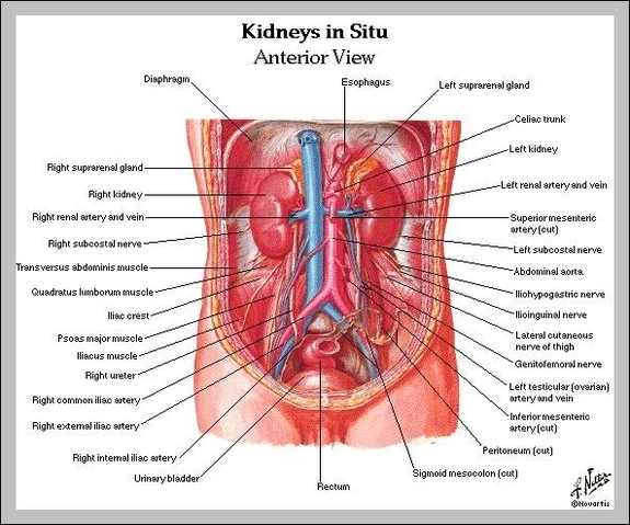
Kidney Human Anatomy: Kidney anatomy includes the cortex, medulla, nephrons, and renal pelvis. These organs filter blood, balance electrolytes, and produce urine.
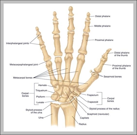
Joints Of The Hand And Wrist: The joints of the hand and wrist include the carpometacarpal joint, metacarpophalangeal joint, and interphalangeal joints, allowing for flexibility and dexterity.
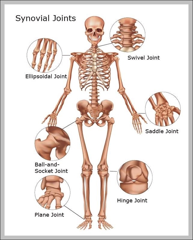
Joints In The Skeletal System: Joints, including hinge, ball-and-socket, pivot, and gliding types, connect bones within the skeletal system and allow for a wide range of movements.
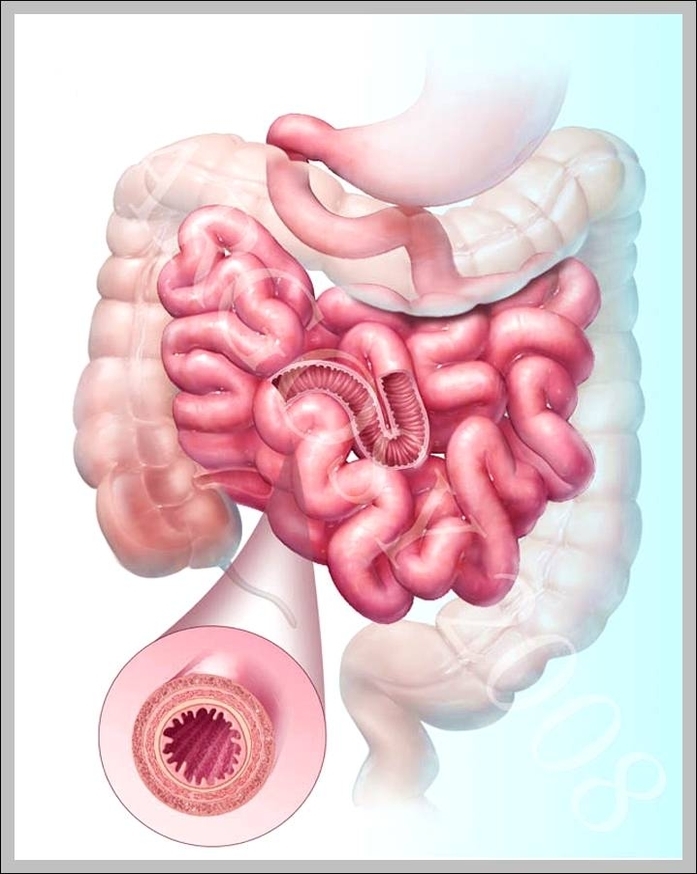
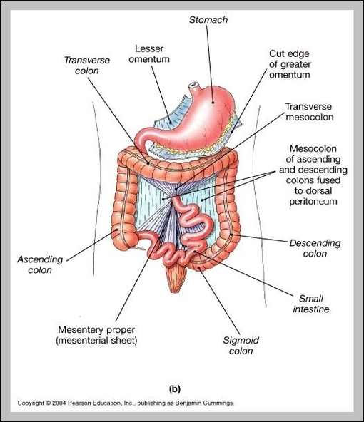
Intestines Image: An intestines image outlines the small intestines coiled structure for nutrient absorption and the large intestines broader path for waste elimination.
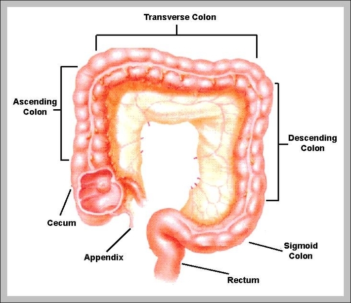
Intestine Length: The small intestine is about 20 feet long, while the large intestine measures around 5 feet, together aiding in digestion, absorption, and waste elimination.
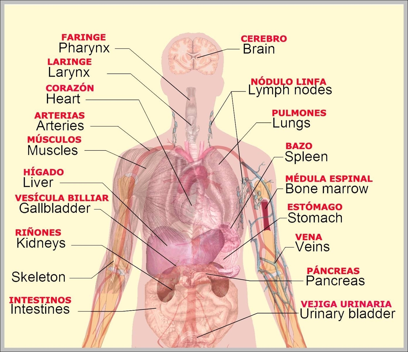
Internal Organs Pictures: Pictures of internal organs display the heart, lungs, liver, stomach, intestines, and kidneys, offering a visual guide to human body systems.
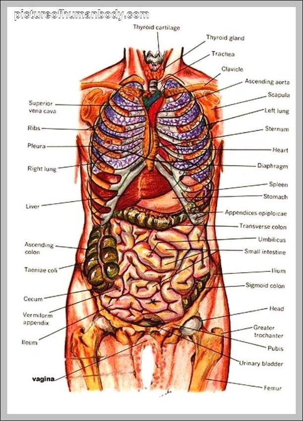
Internal Organs Anatomy: Internal organ anatomy includes vital structures such as the heart, lungs, liver, kidneys, pancreas, and intestines that maintain bodily functions.
Internal Organ Map: An internal organ map visually represents the location of the major organs within the human body. It typically highlights the heart, lungs, liver, kidneys, and digestive organs, helping to understand their placement and function.
Internal Organ Locations: Internal organ locations refer to the positions of organs like the heart, lungs, liver, kidneys, and intestines within the body, often shown in anatomical diagrams.
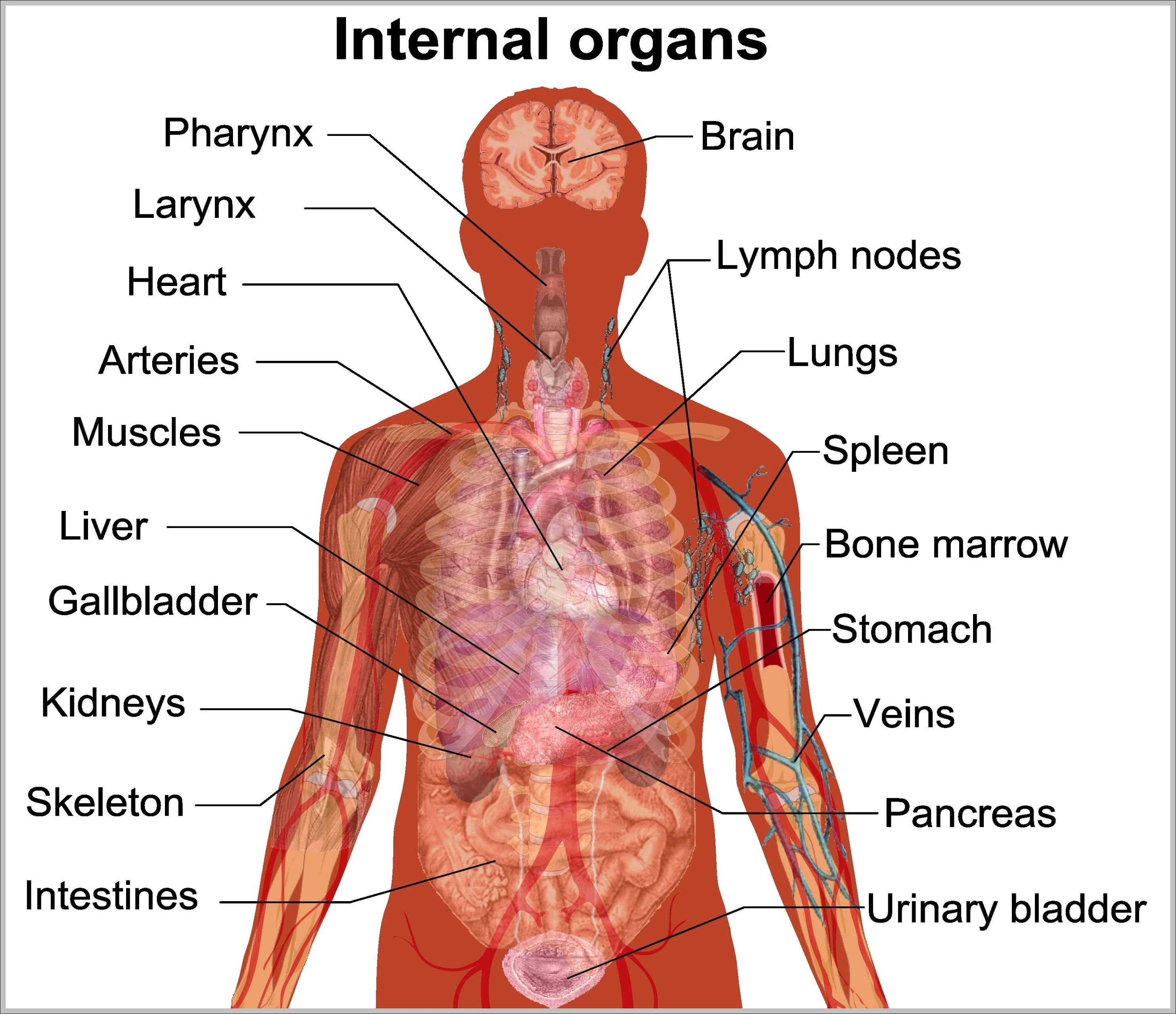
Internal Organ Chart: An internal organ chart labels organs like the heart, lungs, stomach, and intestines, often grouped by system and location for easy reference.