Auditory Ossicles Diagram Image

This human anatomy diagram with labels depicts and explains the details and or parts of the Auditory Ossicles Diagram Image. Human anatomy diagrams and charts show internal organs, body systems, cells, conditions, sickness and symptoms information and/or tips to ensure one lives in good health.

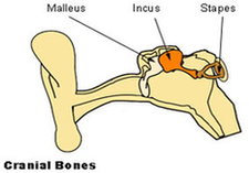
In this article, we’ll discuss the auditory ossicles, namely the malleus, incus, and stapes. Inside of the middle ear are the smallest bones in the body–the auditory ossicles, or ear bones. By definition, these three bones are named after their shape: malleus (“hammer”), incus (anvil), and stapes (stirrup).

By definition, these three bones are named after their shape: malleus (“hammer”), incus (anvil), and stapes (stirrup). During development, the auditory ossicles are the first bones to fully ossify and are mature at the time of birth, where they do not grow afterwards. The ossicles are situated in the middle ear and suspended by ligaments.

The incus (anvil) is the middle auditory ossicle. It is suspended medial to the malleus and lateral to the stapes and joins these ossicles together with synovial joints. It consists of the: body, short limb, long limb/process, and lenticular process.

Auditory Ossicles Diagram Image

Auditory Ossicles Diagram Image


Tags: ,