Peroneal Muscle Image

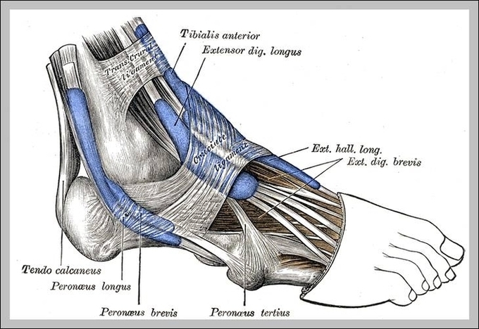
The peroneal muscles are a group of two muscles of the leg that lie within the peroneal compartment located at the lateral fibular region.

The deep peroneal nerve, meanwhile, connects to the muscles of the front of your calf, including tibialis anterior, extensor digitorum longus, and extensor hallucis longus. Those muscles allow you to flex your foot and extend your toes. The common peroneal nerve and its branches also serve sensory functions: 1 

The muscle is considered an extrinsic ankle muscle; it originates in your leg and attaches to your foot and serves to move your ankle. The peroneus longus tendons are held in place near your lateral ankle by the superior peroneal retinaculum, a thick band of tissue. Peroneus longus is a superficial muscle that can easily be seen and palpated.

Peroneal Muscle Image Diagram - Chart - diagrams and charts with labels. This diagram depicts Peroneal Muscle Image

Peroneal Muscle Image