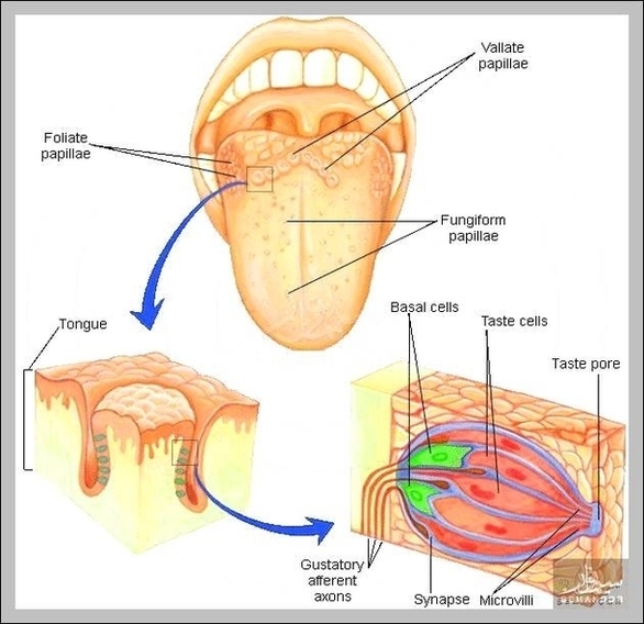
Fungiform Papillae Image
Fungiform papillae, magnified and sectional diagram. The fungiform papillae are club shaped projections on the tongue, generally red in color. They are found on the tip of the tongue, scattered amongst the filiform papillae but are mostly present on the View Diagram Fungiform Papillae Image