Diagram Of Fetal Head Lateral Image

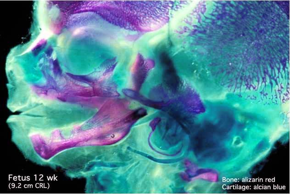
The important structures in the fetal head can be obtained using an axial plane. After obtaining the first set of images from the BPD level, rotation of the transducer will bring the cerebellum and the posterior fossa in to view. 1 caliper is placed outside of the skull bone, and contralateral caliper is placed on the inside of the skull bone

The lower right images demonstrate that angling the fetal head can improve visualisation of the near field lateral ventricle. 1 Near field posterior horn of lateral ventricle 2 Choroid 3 Far field posterior horn of lateral ventricle 4 Cavum Septum Pellucidum (CSP) 5 Corpus callosum (CC)

Since the transverse diameter is greater than the antero-posterior (AP) diameter in the pelvic inlet, the widest circumference of the fetal head descends in a transverse position.

Diagram Of Fetal Head Lateral Image Diagram - Chart - diagrams and charts with labels. This diagram depicts Diagram Of Fetal Head Lateral Image

Diagram Of Fetal Head Lateral Image