Cecum is a part of the digestive system. It comes under the large intestine along with the colon. Basically, the body possesses two types of intestines. The small intestine is attached to the stomach and controls the middle part of the digestion. Stomach To Cecum Watse Diagram Image Diagram - Chart - diagrams and charts with labels. This diagram depicts Stomach To Cecum Watse Diagram Image and explains the details of Stomach To Cecum Watse Diagram Image.
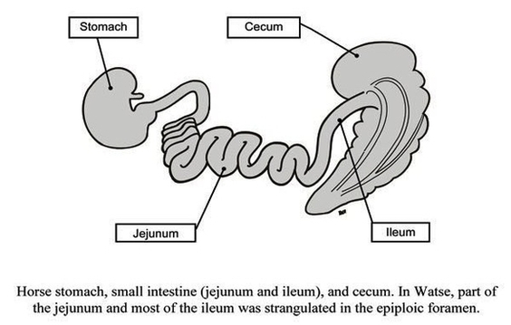
Stomach To Cecum Watse Diagram Image