Picture Of a Knee Cap

This human anatomy diagram with labels depicts and explains the details and or parts of the Picture Of a Knee Cap. Human anatomy diagrams and charts show internal organs, body systems, cells, conditions, sickness and symptoms information and/or tips to ensure one lives in good health.

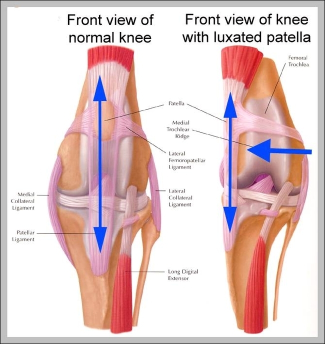
Knee joint: The knee joint has three parts. The thigh bone (the femur) meets the large shin bone (the tibia) to form the main knee joint. This joint has an inner (medial) and an outer (lateral) compartment. The kneecap (the patella) joins the femur to form a third joint, called the patellofemoral joint.

The patella (kneecap) sits over the front of the knee joint. Four major ligaments connect the bones and stabilize the knee joint . In this image, the physician is pointing to the anterior cruciate ligament, or ACL, one of these important ligaments. Inside the knee joint is a smooth cover on the ends of the bone called articular cartilage.

Fracture – a knee bone fracture is also a cause in the swelling of the kneecap. Either bone of the knee joint when gets fractured due to an injury or an accident causes a lot of pain, redness and swelling in the area.

Picture Of a Knee Cap

picture of a knee cap