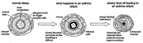
Development of Animal Models Early animal models of asthma were developed in a variety of species (90) and focused on the phenomenon of airways hyperresponsiveness (AHR), defined as excessive bronchoconstriction in response to a standardized challenge. The challenging agent was usually a smooth muscle agonist such as methacholine or histamine. Pb Modelling The Human Ventilation System Asthma Image Diagram - Chart - diagrams and charts with labels. This diagram depicts Pb Modelling The Human Ventilation System Asthma Image and explains the details of Pb Modelling The Human Ventilation System Asthma Image.
Pb Modelling The Human Ventilation System Asthma Image