Ear Anatomy With Labels

This human anatomy diagram with labels depicts and explains the details and or parts of the Ear Anatomy With Labels. Human anatomy diagrams and charts show internal organs, body systems, cells, conditions, sickness and symptoms information and/or tips to ensure one lives in good health.

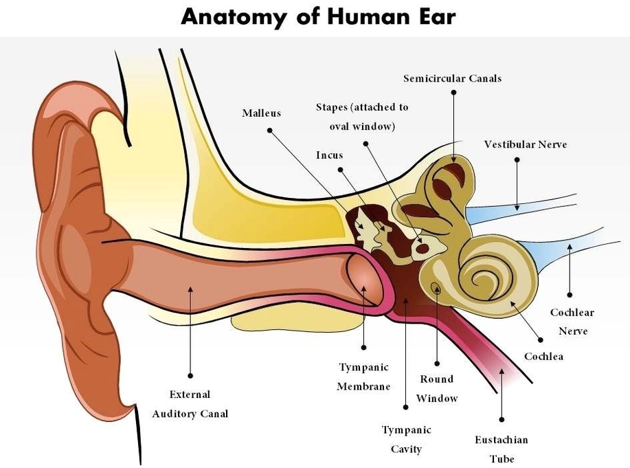
Figure 2. Ear anatomy. The external (outer) ear consists of the auricle, external auditory canal, and eardrum (Figure 1 and 2). The auricle or pinna is a flap of elastic cartilage shaped like the flared end of a trumpet and covered by skin. The rim of the auricle is the helix; the inferior portion is the lobule.

Under the skin the outer one third of the canal is cartilage and inner two thirds is bone. The ear drum is about the size of a dime and is the same size in the new born baby as in the adult. The medical term for the ear drum is the tympanic membrane. The ear drum is a transparent gray membrane.

The ear is made up of three parts: the outer, middle, and inner ear. All three parts of the ear are important for detecting sound by working together to move sound from the outer part through the middle and into the inner part of the ear. Ears also help to maintain balance.

Ear Anatomy With Labels

Ear anatomy with labels


Tags: , , ,