Diverticulitis Diagram Image

This human anatomy diagram with labels depicts and explains the details and or parts of the Diverticulitis Diagram Image. Human anatomy diagrams and charts show internal organs, body systems, cells, conditions, sickness and symptoms information and/or tips to ensure one lives in good health.

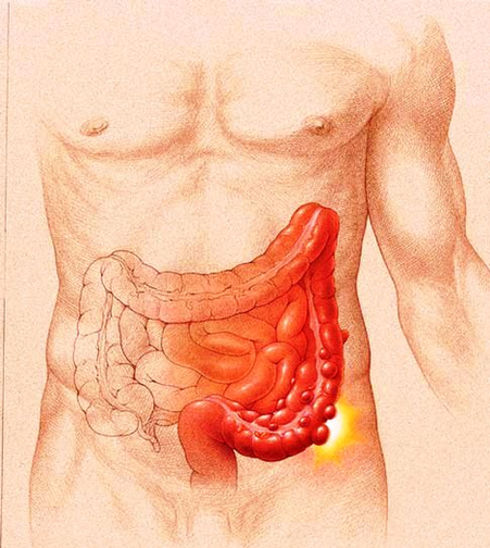
Diverticulosis is a condition where a patient has diverticula in the colon. A patient who suffers the consequences of diverticulosis in the colon is referred to as having diverticular disease. Single-contrast barium enema study in a patient with diverticulitis demonstrates tethering of the sigmoid colon as a result of a diverticular abscess.

Diverticula are pouches that protrude beyond the colon wall due to areas of muscle weakness. Diverticula can be visualized on a barium x-ray, which is an x-ray that is taken after the administration of a barium enema. Problem areas in the colon can be further examined with the help of colonoscopy or flexible sigmoidoscopy.

Diverticulitis results of the inflammation of one of the diverticula. Diverticula in the large intestine. Diverticulitis results of the inflammation of one of these diverticula. The most common sympton is abdominal pain.”n diverticulitis stock pictures, royalty-free photos & images

Diverticulitis Diagram Image

Diverticulitis Diagram Image


Tags: