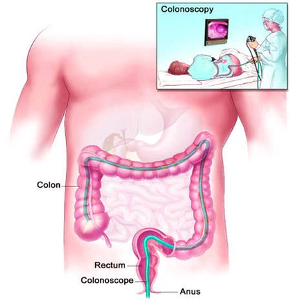
The scope — which is long enough to reach the entire length of your colon — contains a light and a tube (channel) that allows the doctor to pump air or carbon dioxide into your colon. The air or carbon dioxide inflates the colon, which provides a better view of the lining of the colon.
Colonoscopy is one of the most sensitive tests currently available for colon cancer screening. The doctor can view your entire colon and rectum. Abnormal tissue, such as polyps, and tissue samples (biopsies) can be removed through the scope during the exam.
During a virtual colonoscopy, a CT scan produces cross-sectional images of the abdominal allowing the doctor to detect changes or abnormalities in the colon and rectum. To help create clear images, a small tube (catheter) is placed inside your rectum to fill your colon with air or carbon dioxide.
Colonos Screening Diagram Image

