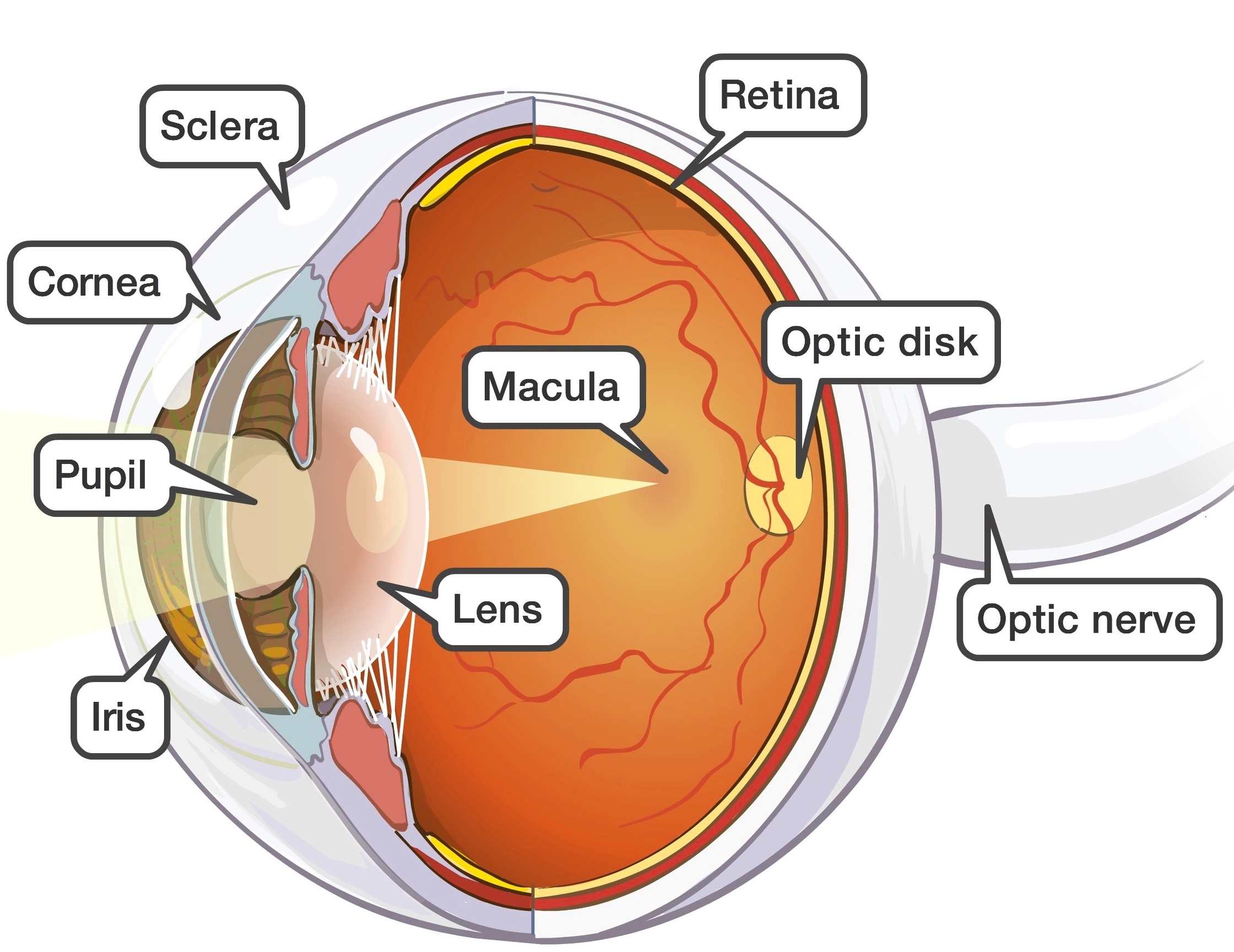
Human Eye Explained
Human Eye Explained Diagram - Chart - diagrams and charts with labels. This diagram depicts Human Eye Explained

Human Anatomy Diagrams
Human Anatomy and Health
Meticulous detail makes this the best 3D heart model ever created. Strip back the layers of the heart and hide or fade individual structures to examine every part. View the heart in action like never before. Set the heart rate to simulate real scenarios, or control the animation to examine every minute movement.
Draw directly on the model, or add Labels to mark out structures for future reference. Change the heart model to show what you need using Tools. Add Growths or Pain to illustrate conditions, or use Cut to access deeper areas of the organ. Cut throught the heart to discover new views and gain deeper understanding.
Get started with a free 3-day trial of Complete Heart on your chosen platform. Access the entire range of features, and begin experiencing the cardiovascular anatomy like never before.
If the kidney concentration is impaired and the patient can achieve a specific gravity of only 1.010, oliguria is present at urine volumes <1000–1500 mL/day. In an attempt to standardize the diagnosis of acute renal failure, the new terminology acute kidney injury (AKI) was coined. Classifying AKI as oliguric or nonoliguric on the basis of daily urine excretion has prognostic value. Oliguria is defined as a daily urine volume of less than 400 mL and has a worse prognosis. Anuria is defined as a urine output of less than 100 mL/day and, if abrupt in onset, suggests bilateral obstruction or catastrophic injury to both kidneys. (Approximately 50-60% of all causes of AKI are nonoliguric.) This lack of a uniform clinical presentation reflects the variable nature of the injury. Classifying AKI as oliguric or nonoliguric on the basis of daily urine excretion has prognostic value. Oliguria is defined as a daily urine volume of less than 400 mL and has a worse prognosis.
Oliguria Acute Kidney Injury Diagram - Chart - diagrams and charts with labels. This diagram depicts Oliguria Acute Kidney Injury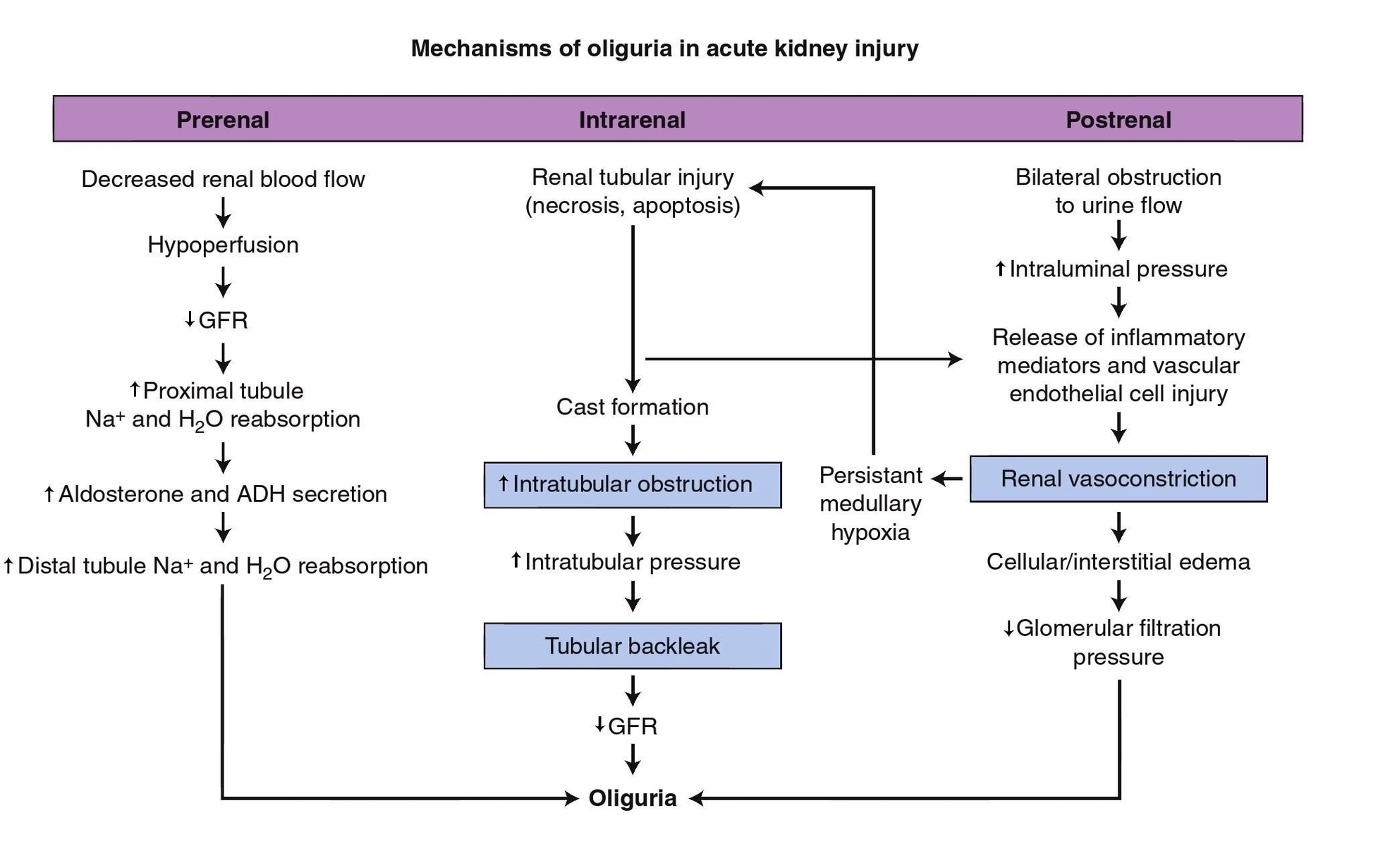
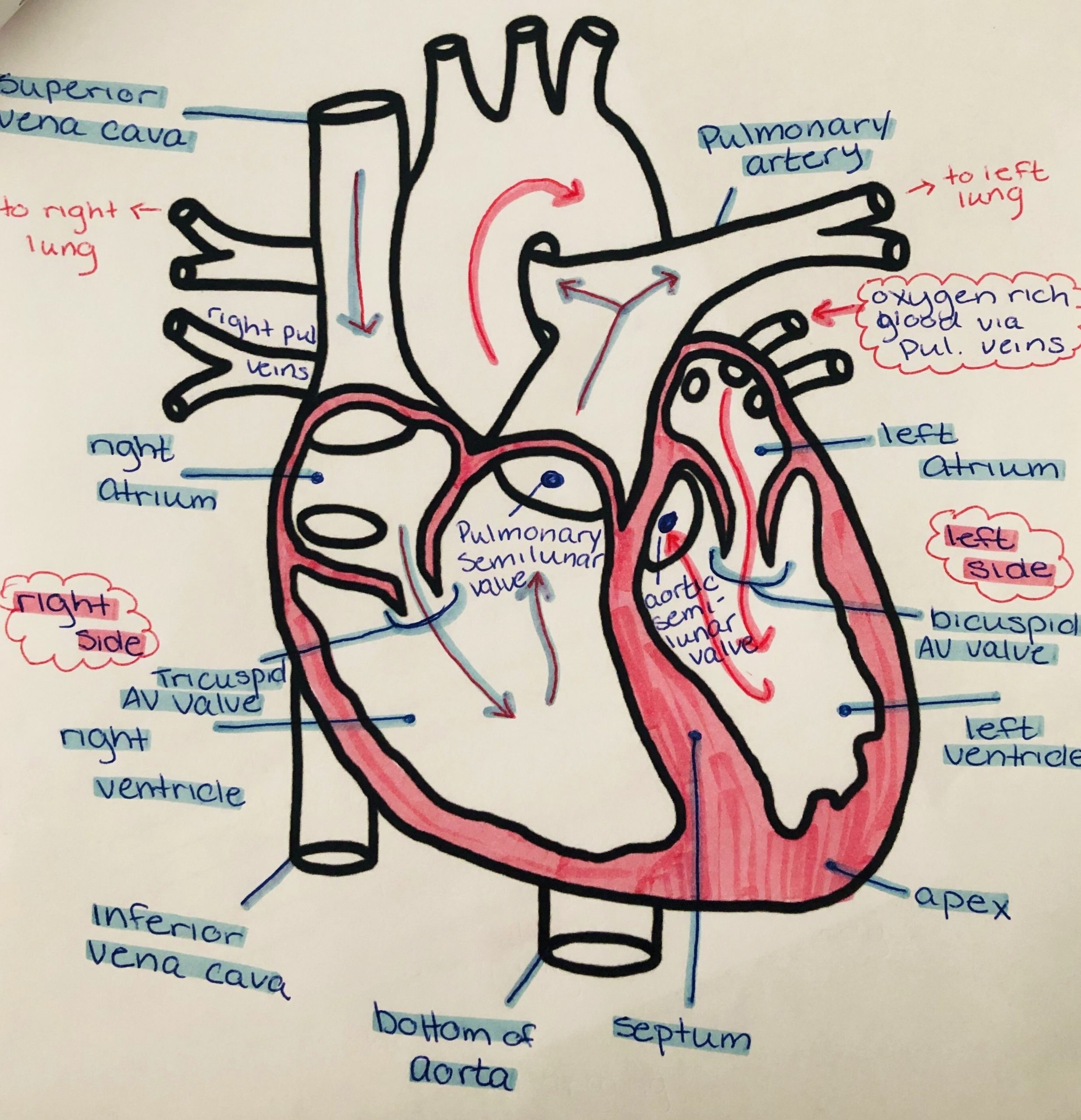
Use your pen or pencil to begin drawing the essential part of your heart diagram, which would look like an open-ended acorn; Draw the image, and angle it to the left around 120 degrees. The critical structure will form the base for ventricles on the left and right;
Drawing a Heart and Arrow. Outline sketch with a circle. Draw another smaller circle overlapping the previous circle. Draw the downward triangle with a little perspective to it. Draw the first cheek. Add the second cheek. Erase the outline sketch and make a new one for the arrow. Always draw the arrow in a slanted way.
To draw a human heart, first draw what looks like the lower half of an acorn that’s missing its cap. This will be the outline of the left and right ventricles. Draw a rounded bump on the top left half of the acorn shape, which will be the right atrium.
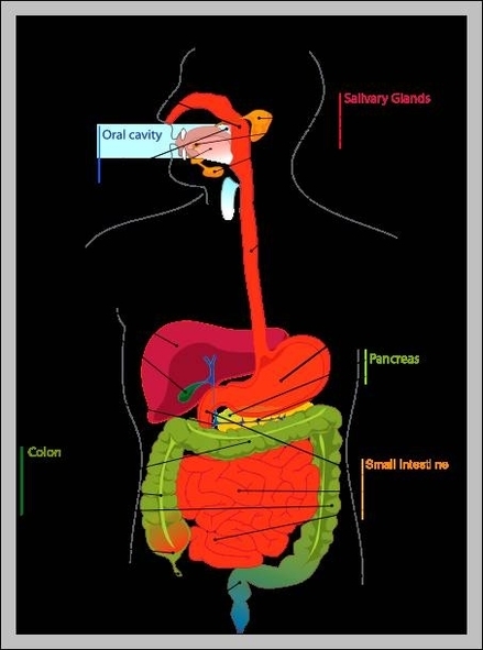
You can find pancreas location in body in the vicinity of other abdominal organs, like liver, gallbladder, spleen, stomach and duodenum. Extending horizontally across the upper left abdomen, it is located behind the stomach.
The pancreas is an abdominal organ that is located behind the stomach and is surrounded by other organs, including the spleen, liver and small intestine. The pancreas is about 6 inches (15.24 centimeters) long, oblong and flat.
The pancreas is located in the abdominal cavity in humans. More specifically, it extends horizontally across the upper left abdomen, it is located behind the stomach. As you can see in the following pancreas diagram and pictures showing other organs as well, it extends to the spleen on the left side and is nestled between the kidneys.
Where Is The Pancreas Located In The Human Body Diagram - Chart - diagrams and charts with labels. This diagram depicts Where Is The Pancreas Located In The Human Body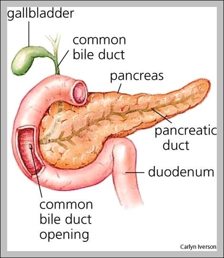
The small intestine is made up of thee sections, including the duodenum, the jejunum and the ileum. On its proximal (near) end, the small intestine—beginning with the duodenum—connects to the stomach. On its distal (far) end, the ileum—the last segment of the small intestine—connects to the large intestine (colon).
Small Intestine. The small intestine is a long, highly convoluted tube in the digestive system that absorbs about 90% of the nutrients from the food we eat. It is given the name “small intestine” because it is only 1 inch in diameter, making it less than half the diameter of the large intestine.
The small intestine (commonly referred to as the small bowel) is a tubular structure/organ that is part of the digestive system. In fact, it is the longest portion of the digestive system, approximately 20 to 25 feet in length.
The Small Intestine Diagram - Chart - diagrams and charts with labels. This diagram depicts The Small Intestine
The main function of the thymus gland is to release thymosin hormone that will stimulate the maturation of T cells. All of our childhood, white blood cells or lymphocytes will come in contact with the thymus gland.
The thymus is a specialized primary lymphoid organ of the immune system. Within the thymus, T cells mature. T cells are critical to the adaptive immune system, where the body adapts specifically to foreign invaders.
Thymus Gland. The thymus gland, despite containing glandular tissue and producing several hormones, is much more closely associated with the immune system than with the endocrine system.
Role Of Thymus Diagram - Chart - diagrams and charts with labels. This diagram depicts Role Of Thymus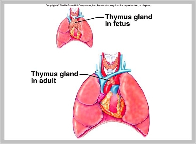
The thyroid gland and parathyroid glands are a group of endocrine glands located in the base of the neck. These glands play a vital role in maintaining the body’s homeostasis by producing hormones that regulate the body’s metabolism and free calcium levels. Variations in thyroid hormones can lead to drastic changes in…
Parathyroids are NOT related to the thyroid (except they are neighbors in the neck). The thyroid gland controls much of your body’s metabolism, but the parathyroid glands control body calcium. They have no relationship except they are neighbors. Parathyroid glands make a hormone, called “Parathyroid Hormone”.
The thyroid gland controls much of your body’s metabolism, but the parathyroid glands control body calcium. They have no relationship except they are neighbors. Parathyroid glands make a hormone, called “Parathyroid Hormone”. Doctors and labs abbreviate Parathyroid Hormone as “PTH”.
Thyroid And Parathyroid Glands Diagram - Chart - diagrams and charts with labels. This diagram depicts Thyroid And Parathyroid Glands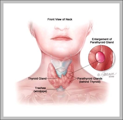
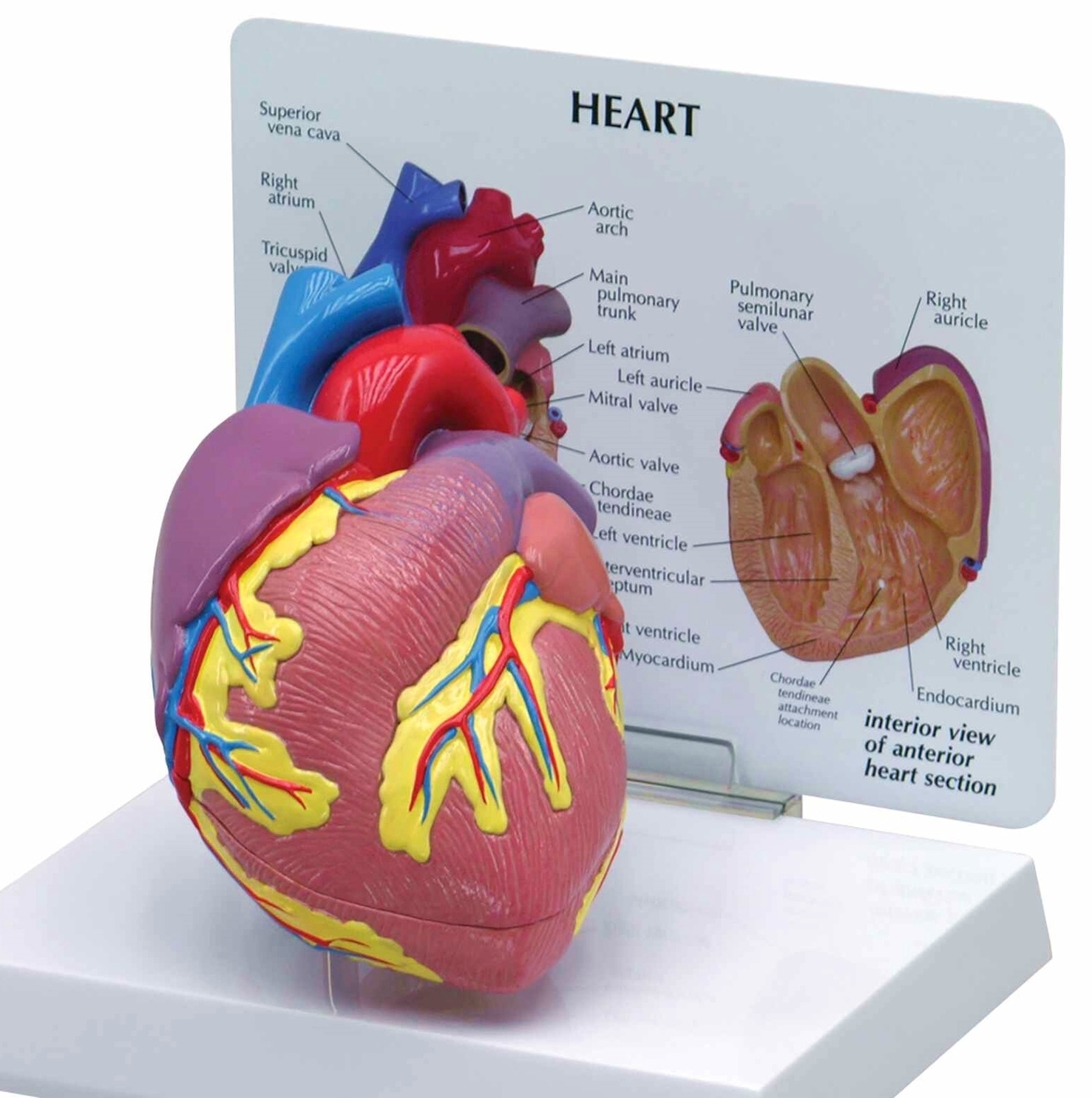
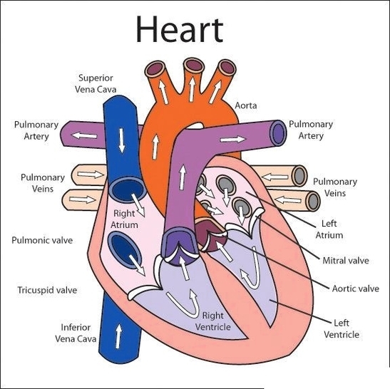
Picture of Heart. It is positioned in the chest behind the sternum (breastbone; in front of the trachea, esophagus, and aorta; and above the diaphragm muscle that separates the chest and abdominal cavities. The normal heart is about the size of a closed fist, and weighs about 10.5 ounces. It is cone-shaped, with the point…
The heart is a mostly hollow, muscular organ composed of cardiac muscles and connective tissue that acts as a pump to distribute blood throughout the body’s tissues.
Picture of Heart. The normal heart is about the size of a closed fist, and weighs about 10.5 ounces. It is cone-shaped, with the point of the cone pointing down to the left. Two-thirds of the heart lies in the left side of the chest with the balance in the right chest.
Pictures Of The Heart Diagram - Chart - diagrams and charts with labels. This diagram depicts Pictures Of The Heart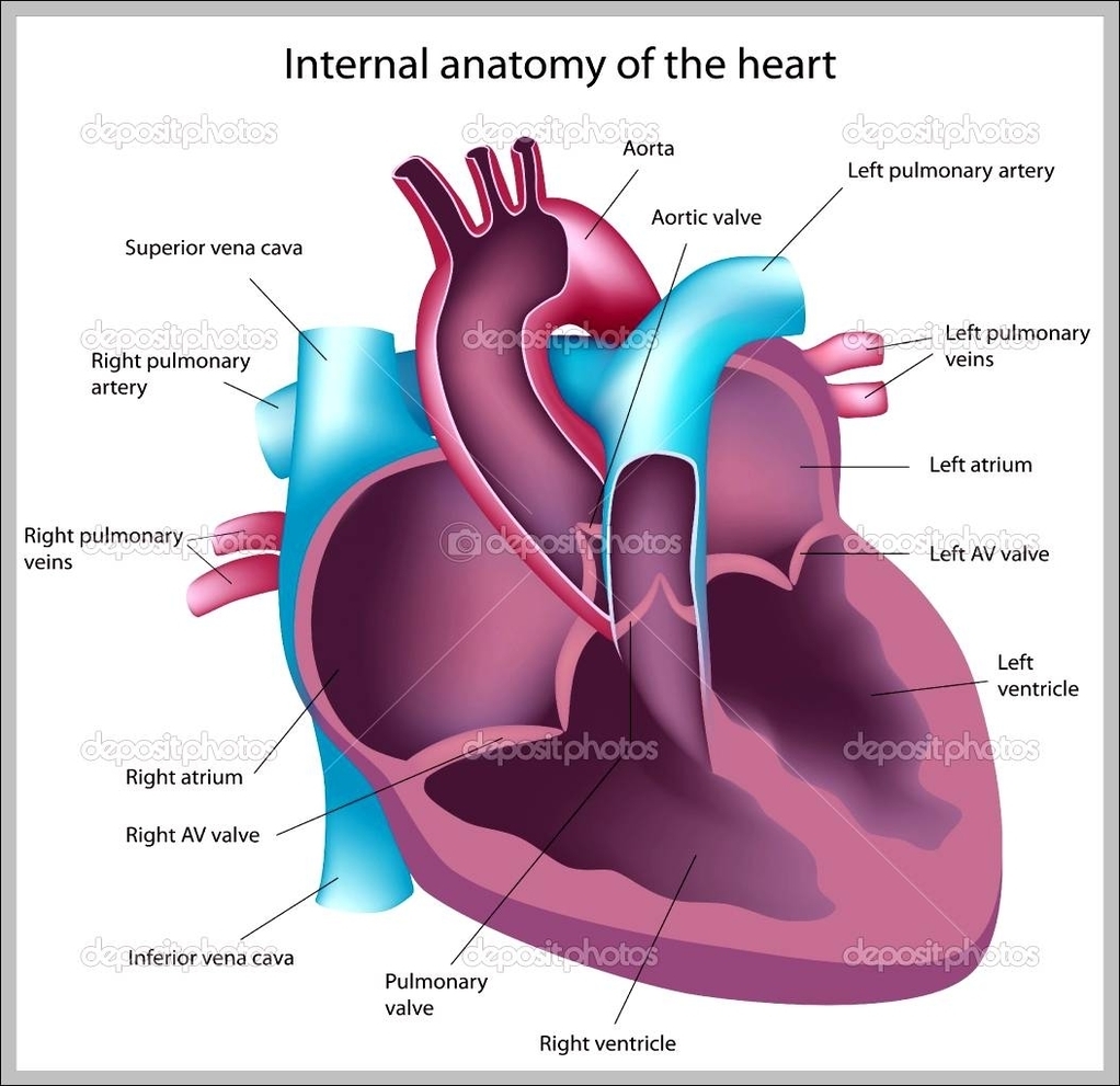
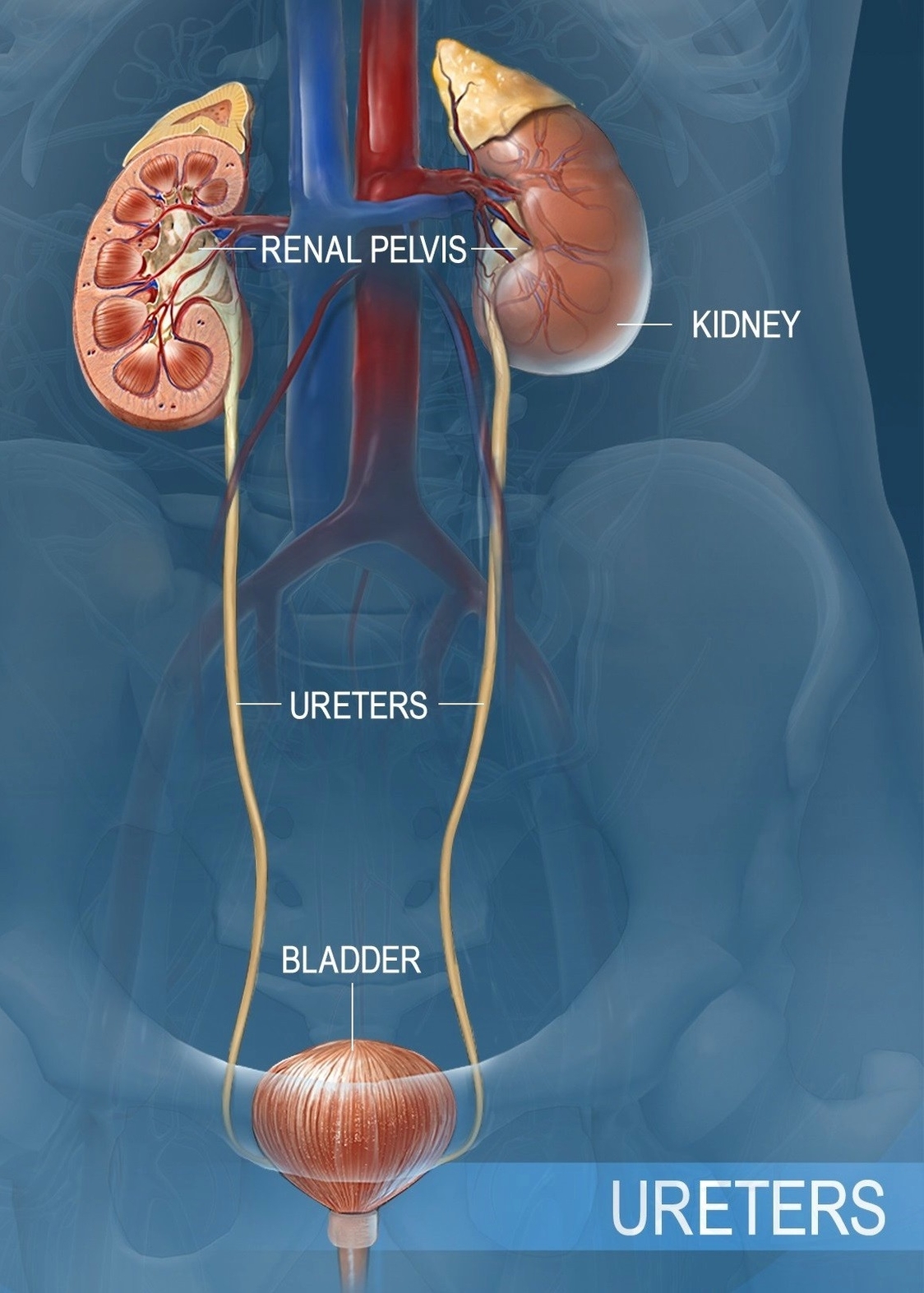
Function. The primary function of the kidneys is to filter the blood and remove excess fluid from it. The kidneys leave just the right amount of salt and other minerals in the blood. The excess amount is filtered from the blood in the form of a waste liquid called urine.
The kidneys are two bean-shaped organs in the renal system. They help the body pass waste as urine. They also help filter blood before sending it back to the heart. The kidneys perform many crucial functions, including: maintaining overall fluid balance.
Urine Formation – Another Vital Function of the Kidney Once the kidneys have filtered the blood and removed the waste products from it, the next step is to get rid of the wastes from the body. Urine formation is the process by which the kidneys prepare waste products, filtered from the blood, for elimination from the body.
What Is The Function Of The Kidney Diagram - Chart - diagrams and charts with labels. This diagram depicts What Is The Function Of The Kidney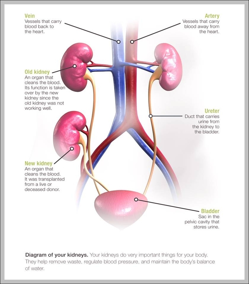
They are located below the rib cage, behind the abdomen, and under the liver. The right kidney sits a bit lower than the left one as the biggest part of the liver is on the right side of the abdomen. Most problems or infections with your kidney cause middle back pain or flank pain. However, the pain is not always felt where the kidneys are located.
The Location of the Kidneys. Your kidneys are two organs about the size of your fist that are shaped like a bean. According to Dr. Charles Patrick Davis on MedicineNet, your kidneys are located just below your rib cage in the back of your abdomen under your liver.
This will help you tell the difference between general back pain and pain that comes from your kidneys. Your kidneys are two organs about the size of your fist that are shaped like a bean. According to Dr. Charles Patrick Davis on MedicineNet, your kidneys are located just below your rib cage in the back of your abdomen under your liver.
Where Is Your Kidney Located Diagram - Chart - diagrams and charts with labels. This diagram depicts Where Is Your Kidney Located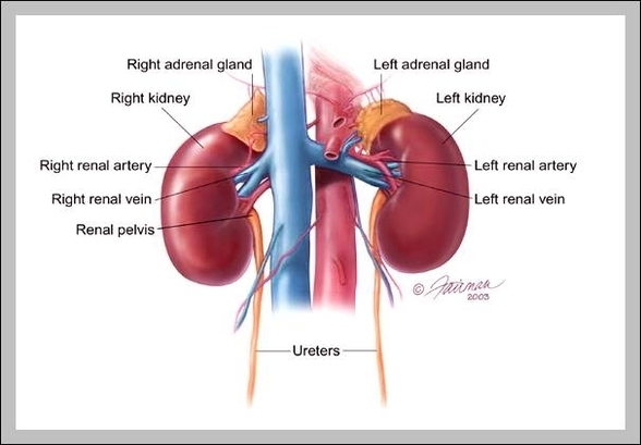
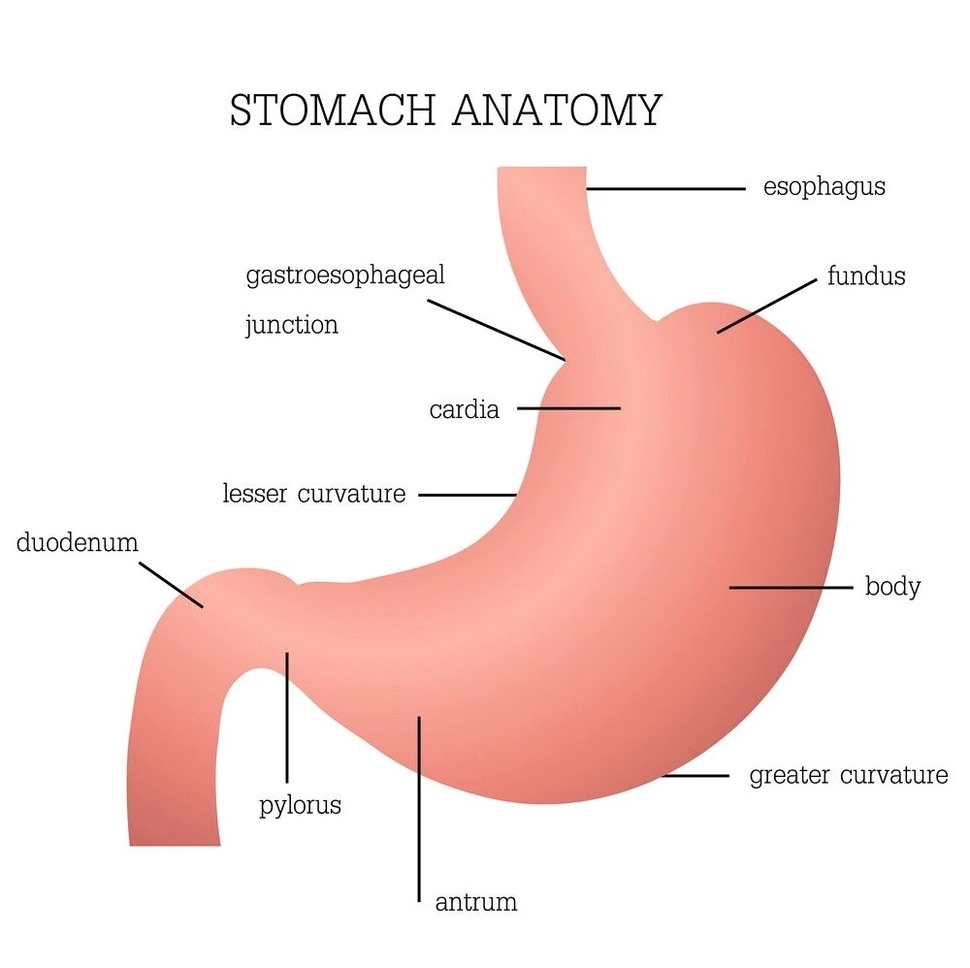
The stomach is an important organ in the digestive system. After food has been chewed in the mouth and swallowed, it enters the stomach via the oesophagus. The stomach produces strong acid. This kills many harmful microorganisms that might have been swallowed along with the food. It also contains special chemicals called enzymes.
The stomach is a muscular, hollow organ in the gastrointestinal tract of humans and many other animals, including several invertebrates. The stomach has a dilated structure and functions as a vital digestive organ. In the digestive system the stomach is involved in the second phase of digestion, following mastication (chewing).
Conditions and diseases. The stomach can have many different conditions and diseases that can cause pain, discomfort, digestion problems and even death. One of the most common stomach problems is upset stomach or dyspepsia.
What Does The Stomach Do Diagram - Chart - diagrams and charts with labels. This diagram depicts What Does The Stomach Do