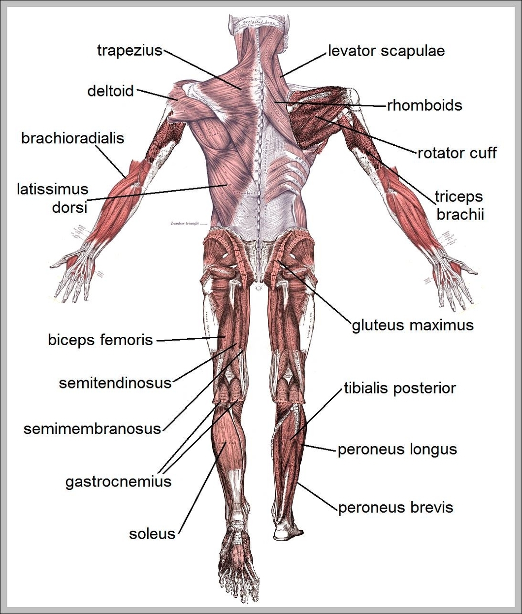
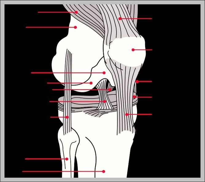
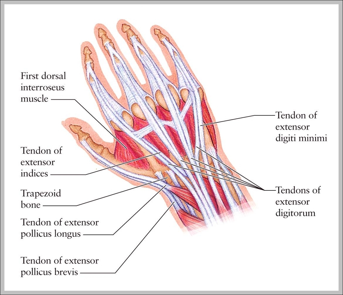
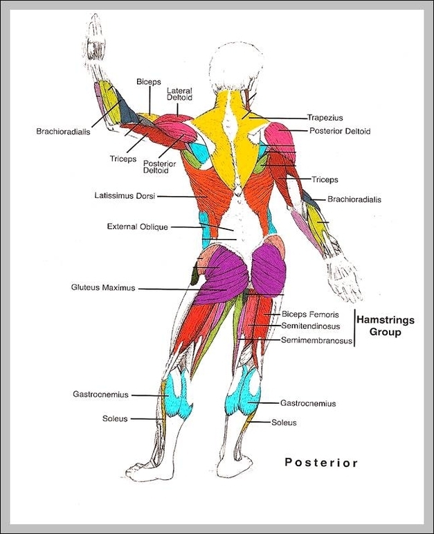
Human Muscle Structure Diagram: A human muscle structure diagram illustrates the layers of muscle tissue, including skeletal muscles, tendons, and the connection to bones for movement.
Human Muscles Labeled Diagram: A human muscles labeled diagram shows the major muscles of the human body, such as the deltoid, biceps, and quadriceps, with labels for educational purposes.
Human Muscles Labeled: A labeled diagram of human muscles displays major muscle groups, such as the biceps, quadriceps, hamstrings, and pectorals, which are essential for movement, stability, and overall body function.
Human Male Body: The human male body includes the male reproductive system, muscular and skeletal structures, and organ systems necessary for growth, function, and reproduction.
Human Liver Location Anatomy: The liver is located in the upper right portion of the abdomen, beneath the diaphragm and above the stomach, and is key to metabolism and detoxification.
Human Liver Function: The liver processes nutrients, detoxifies harmful substances, produces bile, and stores energy, playing a central role in metabolism and digestion.
Human Leg Muscle Diagram: This diagram highlights key leg muscles such as the quadriceps, hamstrings, calf muscles, and glutes. These muscles enable walking, running, jumping, and posture support.
Human Knee Anatomy Diagram: This diagram shows the knees internal components including ligaments, cartilage, synovial membrane, and bones, crucial for load-bearing and mobility.
Human Knee Anatomy Diagram: A human knee anatomy diagram highlights the bones, ligaments, cartilage, and tendons that make up this complex joint, including the femur, tibia, patella, ACL, and menisci.
Human Joints Diagram: This diagram shows joints like ball-and-socket (shoulder, hip), hinge (elbow, knee), and pivot (neck), allowing various movement ranges.
Human Internal Organs Pictures: Images of human internal organs are used for educational and medical reference to identify organs like the brain, heart, lungs, kidneys, and digestive structures. These visuals aid in understanding function and diagnosing conditions.
Human Internal Organ Diagram: A diagram of internal organs reveals the heart, lungs, liver, kidneys, stomach, intestines, and reproductive organs positioned within the torso and pelvic regions.
Human Integumentary System: This system includes skin, hair, nails, and glands, serving as a protective barrier and regulating temperature and sensory input.
Human Heart With Labels: A diagram of the human heart with labels shows the four chambers of the heart, valves, and major blood vessels, illustrating how blood flows through the cardiovascular system.
Human Heart Pictures For Kids: Heart pictures for kids present simplified visuals of the heart, showing its shape, location, and function in a way thats easy to understand and ideal for early anatomy education.
Human Heart Location: The human heart is located slightly left of the center in the chest cavity, behind the sternum and between the lungs, protected by the ribcage.
Human Heart In Body: The human heart is located in the center of the chest, slightly to the left, nestled between the lungs and above the diaphragm, where it pumps blood throughout the circulatory system.
Human Heart Anatomy For Kids: Human heart anatomy for kids is simplified to explain how the heart works in pumping blood throughout the body, with easy-to-understand visuals and descriptions for children.
Human Hand Muscles And Tendons: The hand contains intricate muscles and tendons, including the flexor and extensor groups, that enable precise movements and grip.
Human Femur Bone Picture: A femur image displays the largest bone in the body, showing features like the head, neck, shaft, and condyles, which connect to the pelvis and knee.