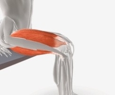
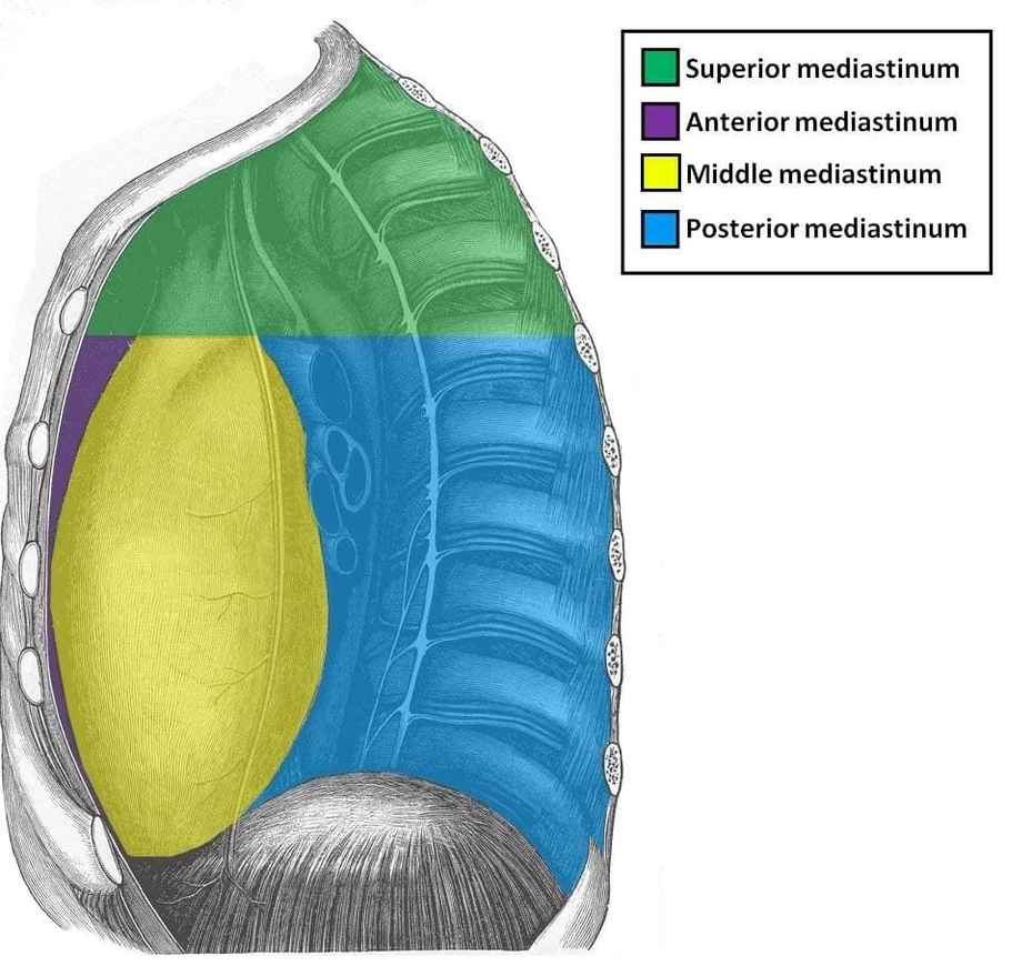
Borders of the Femoral Triangle
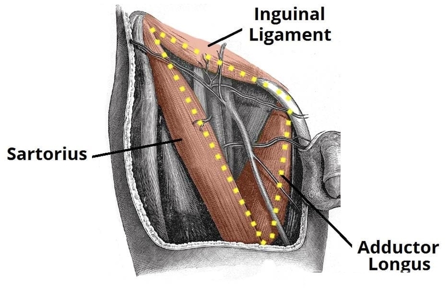
The femoral triangle is an anatomical region in the anterior thigh bounded by the sartorius, adductor longus, and inguinal ligament. It contains key structures including the femoral nerve, artery, vein, and lymphatics. Understanding the borders and contents of the femoral View Diagram Borders of the Femoral Triangle