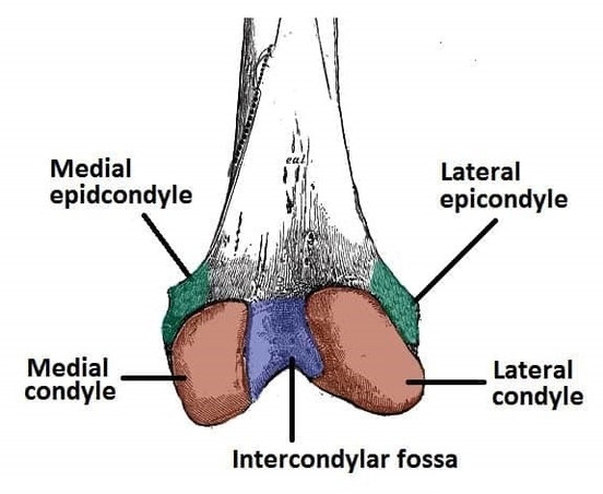
The posterior surface of the distal femur includes the condyles, intercondylar fossa, popliteal surface, and articular facets for the tibia and patella. These structures articulate with the tibial plateau and patella, contributing to knee joint stability, flexion, and extension. Knowledge of distal femur anatomy is essential in orthopedic surgery, fracture fixation, and knee replacement procedures. Understanding bony landmarks, attachment points for ligaments, and relationships to surrounding muscles ensures proper alignment, restoration of joint function, and prevention of post-operative complications. Accurate anatomical orientation is critical for safe interventions and successful rehabilitation of knee injuries. Posterior Surface of the Distal Portion of the Femur Diagram - Chart - diagrams and charts with labels. This diagram depicts Posterior Surface of the Distal Portion of the Femur and explains the details of Posterior Surface of the Distal Portion of the Femur.
Posterior Surface of the Distal Portion of the Femur