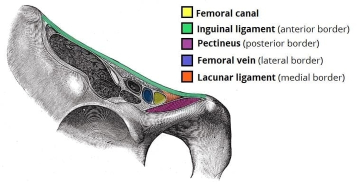
The femoral canal is a small, clinically important space within the femoral triangle and is bounded anteriorly by the inguinal ligament, posteriorly by the pectineal ligament, medially by the lacunar ligament, and laterally by the femoral vein. It contains lymphatic vessels and loose connective tissue, providing space for the femoral vein to expand. Its narrow shape makes it a potential site for femoral hernias, which occur when abdominal contents protrude into the canal. Understanding these borders is essential for surgical repair and for distinguishing femoral hernias from inguinal ones. Borders of the Femoral Canal Diagram - Chart - diagrams and charts with labels. This diagram depicts Borders of the Femoral Canal and explains the details of Borders of the Femoral Canal.
Borders of the Femoral Canal