Week Embryo Brain Image

This human anatomy diagram with labels depicts and explains the details and or parts of the Week Embryo Brain Image. Human anatomy diagrams and charts show internal organs, body systems, cells, conditions, sickness and symptoms information and/or tips to ensure one lives in good health.

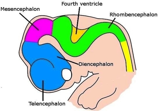
From the time the neural tube closes, around week 7, the brain will grow at a rate of 250,000 neurons per minute for the next 21 weeks. Ultrasounds can reveal the embryo moving as early as 6 weeks after conception (or 8 pregnancy weeks), detecting the electrical impulses that govern movement and indicating that the brain is beginning to function.

By about week 10, the brain is a small, smooth structure that looks a whole lot more like the brain you’re used to (minus the folds that make up the various brain regions, which develop later in pregnancy). When does a fetus have brain activity? The first synapses in baby’s spinal cord form during week 7 of pregnancy.

At just six weeks, the embryo’s brain and nervous system begin to develop, although the complex parts of the brain continue to grow and develop through the end of pregnancy, with development ending around the age of 25.

Week Embryo Brain Image

Week Embryo Brain Image


Tags: , ,