Tubular Organ Diagram Small Image

This human anatomy diagram with labels depicts and explains the details and or parts of the Tubular Organ Diagram Small Image. Human anatomy diagrams and charts show internal organs, body systems, cells, conditions, sickness and symptoms information and/or tips to ensure one lives in good health.

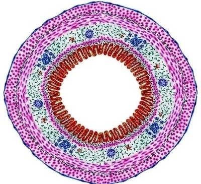
You know esophagus is the best organ that have all the layers of a tubular organ. I am going to describe the different layers from esophagus histology slide. The mucosa of esophagus consists of lamina epithelium, lamina propria and lamina muscularis.

5,814 male human anatomy diagram stock photos and images available, or start a new search to explore more stock photos and images. Male silhouette (contour) on white background, vector. Male, female and children’s silhouette on white background,…

The microscopic images of the urinary bladder and the labeled diagram might help you easily identify the slide under the light microscope. Most characteristic features of the urinary bladder slide are found in the tunica mucosa layer, lining epithelium, and tunica muscularis layer.

Tubular Organ Diagram Small Image

Tubular Organ Diagram Small Image


Tags: , ,