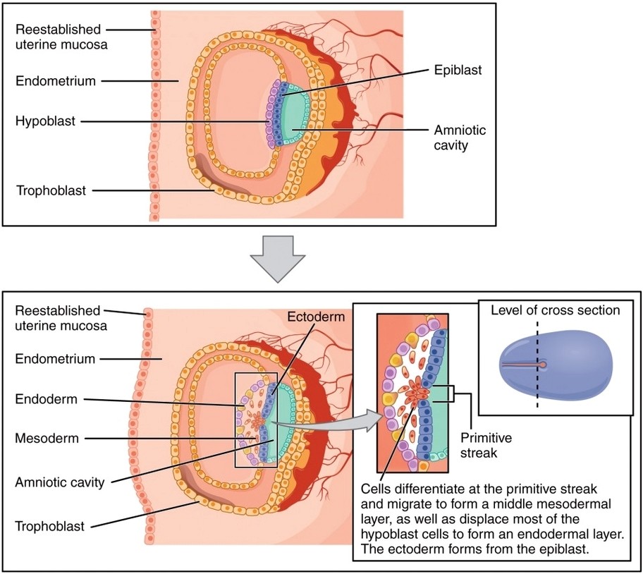
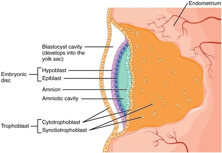
Embryonic Disc Amniotic Cavity Yolk Sac: By the second week of development, the embryonic disc forms between the amniotic cavity above and the yolk sac below, setting the stage for tissue differentiation and body axis formation.
Size of Uterus Throughout Pregnancy: The uterus expands dramatically during pregnancy, growing from a small pelvic organ to a structure that can reach the rib cage to accommodate fetal development.
Photo of Embryo: An embryo photo shows the early stages of human development, typically with visible somites, neural tube, limb buds, and heart bulge around week 46.
Fetal Circulation: Fetal circulation features shunts like the ductus venosus, foramen ovale, and ductus arteriosus that divert blood away from the liver and lungs, directing it efficiently between the heart and placenta.
Photo of Placenta: A placenta photo typically shows the fetal side with vessels radiating from the umbilical cord and the maternal side with a spongy, vascular appearance that interfaces with the uterus.
Germ Layers: The three primary germ layersectoderm, mesoderm, and endodermform during gastrulation and give rise to all tissues and organs in the developing embryo.