Shoulder Diagram

This human anatomy diagram with labels depicts and explains the details and or parts of the Shoulder Diagram. Human anatomy diagrams and charts show internal organs, body systems, cells, conditions, sickness and symptoms information and/or tips to ensure one lives in good health.

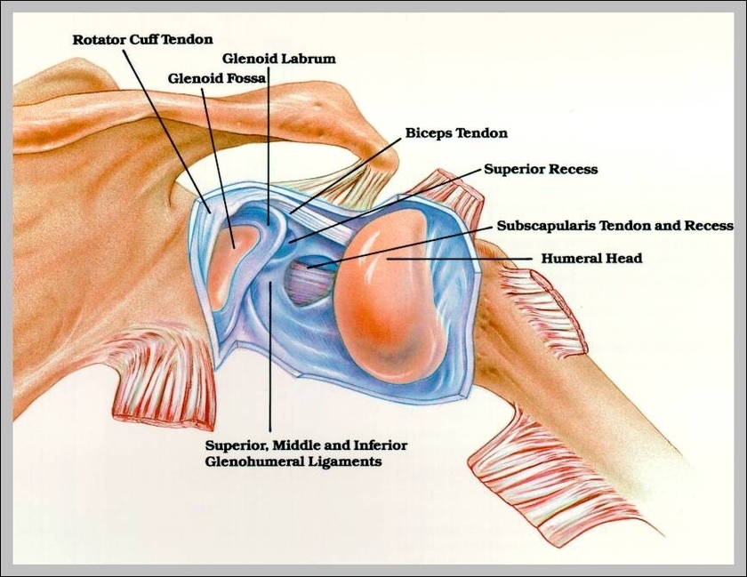
Toggle Anatomy System. Surrounding the rotator cuff muscles are many groups of muscles that work together to produce the various movements of the shoulder. Located superior to the shoulder joint, the deltoid muscle works with the supraspinatus to abduct the arm at the shoulder. On the anterior side of the shoulder, the coracobrachialis,…

The shoulder has several other important structures: The rotator cuff is a collection of muscles and tendons that surround the shoulder, giving it support and allowing a wide range of motion. The bursa is a small sac of fluid that cushions and protects the tendons of the rotator cuff.

It causes pain in the area just outside the joint. Common types of tendinitis include pitcher’s and swimmer’s shoulder. Bursitis is when the bursa (a small sac filled with fluid that protects your rotator cuff) gets irritated.

Shoulder Diagram

shoulder diagram