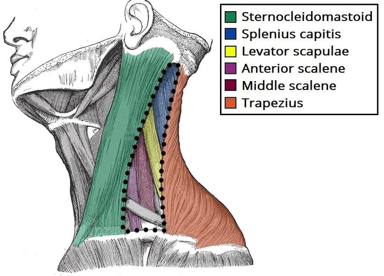
The posterior triangle of the neck is bounded anteriorly by the sternocleidomastoid, posteriorly by the trapezius, and inferiorly by the clavicle. The floor is formed by several muscles including the splenius capitis, levator scapulae, and scalene muscles. This space contains important neurovascular structures such as the accessory nerve, branches of the cervical plexus, and portions of the subclavian vessels. The triangle is clinically significant for surgical access to the neck, lymph node examination, and procedures such as central venous catheterization. Understanding its boundaries and floor muscles is essential to avoid injury to these vital structures. Posterior Triangle of the Neck Borders and Muscles of the Floor Diagram - Chart - diagrams and charts with labels. This diagram depicts Posterior Triangle of the Neck Borders and Muscles of the Floor and explains the details of Posterior Triangle of the Neck Borders and Muscles of the Floor.
Posterior Triangle of the Neck Borders and Muscles of the Floor