Embryology Constructing The Organism Image

This human anatomy diagram with labels depicts and explains the details and or parts of the Embryology Constructing The Organism Image. Human anatomy diagrams and charts show internal organs, body systems, cells, conditions, sickness and symptoms information and/or tips to ensure one lives in good health.

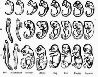
Embryo The embryo model is about 4 weeks old, and mounted on a stand. Embryonic Development The model includes 8 parts showing the uterus with embryo and fetus from the first to the seventh month pregnancy. The very latest technology will involve 3D printed models that will allow researchers, educators and students to make their very own models.

The models include whole surface views as well as detailed views of embryo internal structures. The collection consists of 64 models and approximately 200,000 serial sections of human embryos.

Embryonic Development The model includes 8 parts showing the uterus with embryo and fetus from the first to the seventh month pregnancy. The very latest technology will involve 3D printed models that will allow researchers, educators and students to make their very own models.

Embryology Constructing The Organism Image

Embryology Constructing The Organism Image


Tags: , ,