Ligaments of the Elbow Joint Lateral and Medial Aspect
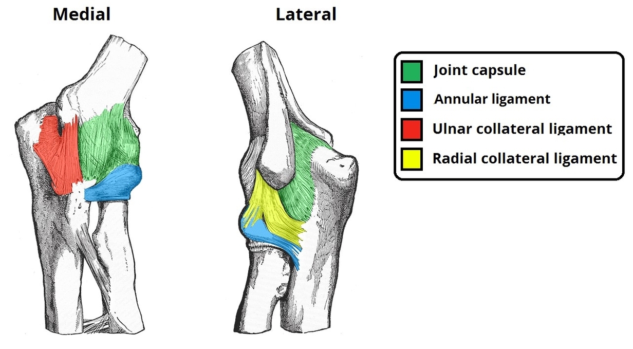
The elbow joint ligaments, including medial (ulnar collateral) and lateral (radial collateral) ligaments, provide stability against varus and valgus stress. They connect the humerus to the ulna and radius, supporting the joint during flexion, extension, and rotational movements. Injury can View Diagram Ligaments of the Elbow Joint Lateral and Medial Aspect