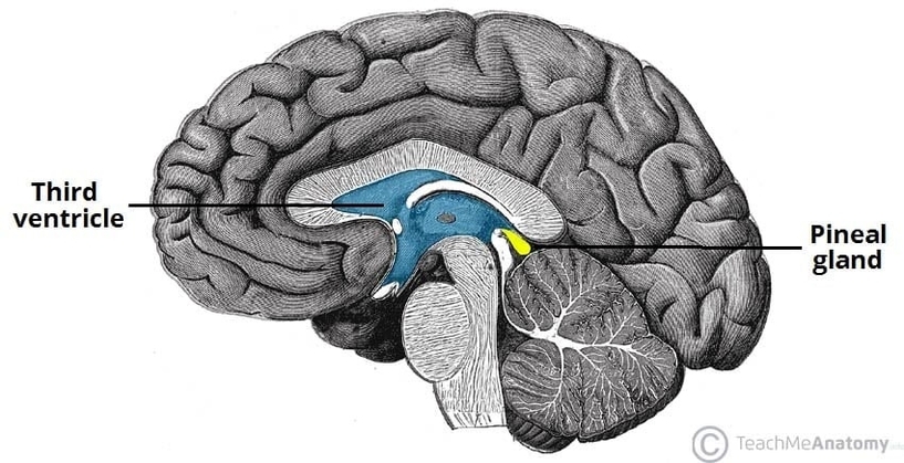
The pineal gland is a small endocrine structure located deep within the brain. This anatomy diagram of the anatomical position of the pineal gland shows its placement in the midline, between the two cerebral hemispheres, within the epithalamus near the third ventricle. It lies posterior to the thalamus and above the superior colliculi. The diagram highlights its role in melatonin secretion and regulation of circadian rhythms. Understanding its anatomical position is important in neuroanatomy and imaging interpretation, particularly when assessing pineal tumors or calcifications. This detailed visual guide supports both anatomical study and clinical neurology learning. Anatomical Position of the Pineal Gland Diagram - Chart - diagrams and charts with labels. This diagram depicts Anatomical Position of the Pineal Gland and explains the details of Anatomical Position of the Pineal Gland.
Anatomical Position of the Pineal Gland