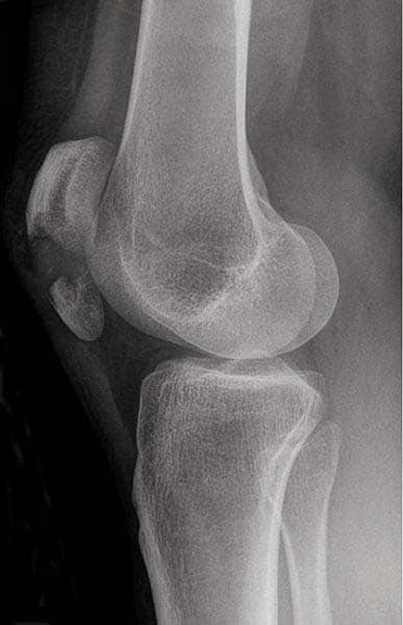
Radiographs of a fractured patella reveal the location, type, and displacement of the fracture. The patella serves as a sesamoid bone within the quadriceps tendon, enhancing knee extension leverage. Understanding patellar anatomy is critical in orthopedics, emergency care, and physiotherapy. Accurate assessment of X-rays informs treatment decisions such as immobilization, surgical fixation, or rehabilitation. Knowledge of surrounding structures, including the quadriceps tendon, patellar ligament, and femoral condyles, ensures safe intervention, restores knee function, and prevents complications like malalignment or impaired mobility. It also helps in post-injury recovery planning, maintaining strength, and preserving the range of motion in the knee joint. Radiograph of Fractured Patella Diagram - Chart - diagrams and charts with labels. This diagram depicts Radiograph of Fractured Patella and explains the details of Radiograph of Fractured Patella.
Radiograph of Fractured Patella