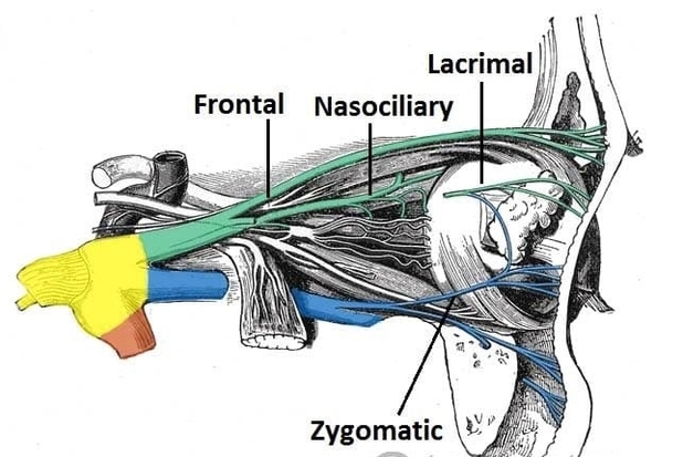
The ophthalmic nerve is the first division of the trigeminal nerve (cranial nerve V1) and provides sensory innervation to the upper face and orbit. This anatomy diagram of the anatomical course of the ophthalmic nerve illustrates its origin from the trigeminal ganglion, passage through the cavernous sinus, and entry into the orbit via the superior orbital fissure. It highlights its three main branches: the frontal, lacrimal, and nasociliary nerves. The diagram clearly demonstrates sensory distribution to the forehead, scalp, upper eyelid, cornea, and dorsum of the nose. Understanding this pathway is essential for diagnosing trigeminal neuralgia, corneal reflex abnormalities, and orbital trauma. This visual resource supports students and clinicians in mastering detailed head and neck neuroanatomy. Anatomical Course of the Ophthalmic Nerve Diagram - Chart - diagrams and charts with labels. This diagram depicts Anatomical Course of the Ophthalmic Nerve and explains the details of Anatomical Course of the Ophthalmic Nerve.
Anatomical Course of the Ophthalmic Nerve