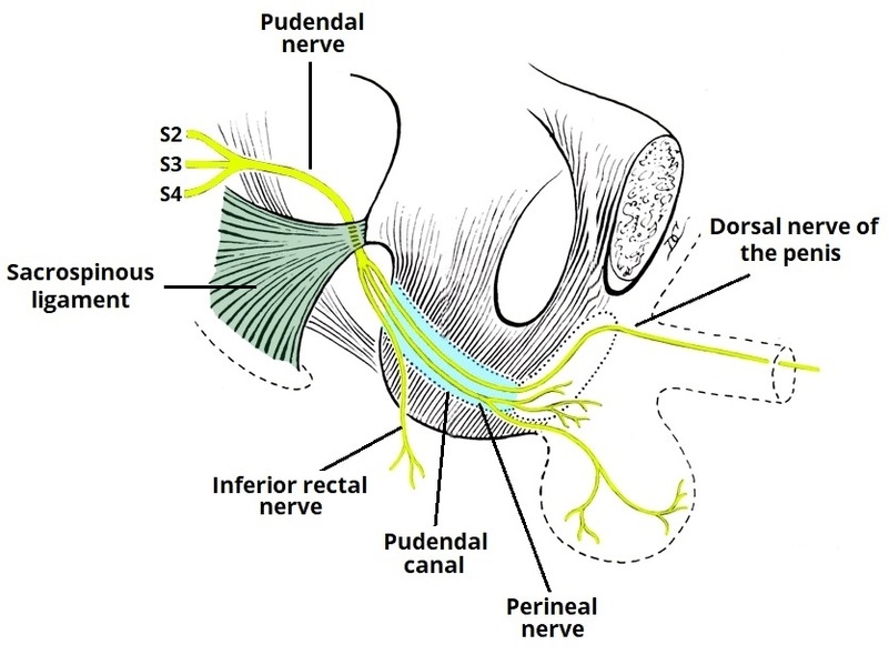
The pudendal nerve is the primary somatic nerve of the perineum, providing both motor and sensory innervation. This anatomy diagram of the anatomical course and branches of the pudendal nerve traces its origin from sacral nerve roots S2S4, its exit through the greater sciatic foramen, and re-entry through the lesser sciatic foramen into the pudendal canal. It highlights key branches including the inferior rectal nerve, perineal nerve, and dorsal nerve of the penis or clitoris. The diagram helps clarify relationships to pelvic muscles, ligaments, and vascular structures. Understanding this course is essential in obstetrics, urology, and the management of pudendal neuralgia. This visual resource supports clinical learning and procedural safety during pelvic surgeries and nerve blocks. Anatomical Course and Branches of the Pudendal Nerve Diagram - Chart - diagrams and charts with labels. This diagram depicts Anatomical Course and Branches of the Pudendal Nerve and explains the details of Anatomical Course and Branches of the Pudendal Nerve.
Anatomical Course and Branches of the Pudendal Nerve