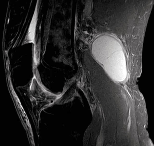
The Bakers cyst appears as a fluid-filled swelling in the popliteal fossa, often secondary to knee joint pathology such as meniscal tears or arthritis. MRI imaging identifies its size, location, and relationship to surrounding structures. Knowledge of Bakers cyst anatomy is essential for orthopedic surgeons, radiologists, and medical students in diagnosis, treatment planning, and aspiration or surgical management. Understanding cyst characteristics aids in differentiating it from tumors, vascular anomalies, or other cystic lesions in the posterior knee. MRI Scan of a Bakers Cyst Diagram - Chart - diagrams and charts with labels. This diagram depicts MRI Scan of a Bakers Cyst and explains the details of MRI Scan of a Bakers Cyst.
MRI Scan of a Bakers Cyst