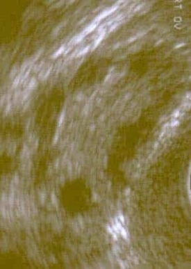
Polycystic ovary syndrome on ultrasound typically shows enlarged ovaries with multiple small follicles arranged around the periphery, often described as a string of pearls. These follicles represent arrested development due to hormonal imbalance, particularly elevated androgens and disrupted ovulatory cycles. The ovarian stroma may appear thickened, reflecting increased hormone production. Ultrasound findings alone are not diagnostic, but they support clinical features such as irregular periods, acne, and insulin resistance. Identifying these sonographic patterns helps guide evaluation and management of the condition. Ultrasound Polycystic Ovary Syndrome Diagram - Chart - diagrams and charts with labels. This diagram depicts Ultrasound Polycystic Ovary Syndrome and explains the details of Ultrasound Polycystic Ovary Syndrome.
Ultrasound Polycystic Ovary Syndrome