The Endocrine System Diagram

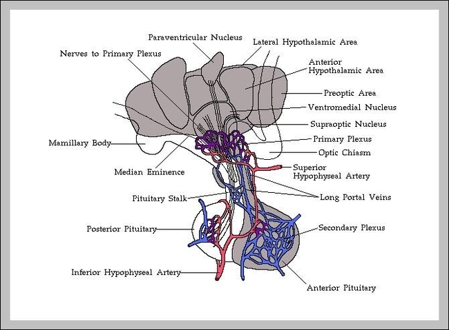
Endocrine System Diagram The endocrine system consists of glands that are found all over the body, which help you to produce hormones. The endocrine system is a collection of glands that secrete various chemicals called hormones. The steroid hormones are derived from cholesterol after a series of biochemical reactions.

This organ connects your endocrine system with your nervous system. Its main job is to tell your pituitary gland to start or stop making hormones. Pituitary gland. This is your endocrine system’s master gland. It uses information it gets from your brain to tell other glands in your body what to do.

The glands of the endocrine system include: Hypothalamus. While some people don’t consider it a gland, the hypothalamus produces multiple hormones that control the pituitary gland. It’s also involved in regulating many functions, including sleep-wake cycles, body temperature, and appetite.

The Endocrine System Diagram Diagram - Chart - diagrams and charts with labels. This diagram depicts The Endocrine System Diagram

the endocrine system diagram

Pics Of Muscular System

Muscular System Parts. 1 Skeletal Muscle. Striated muscle, or Skeletal muscle, is the tissue most commonly associated with the muscular system. This type of muscle attaches to … 2 Visceral Muscle. 3 Cardiac Muscle.

The muscular system is responsible for the movement of the human body. Attached to the bones of the skeletal system are about 700 named muscles that make up roughly half of a person’s body weight.

Muscular System Physiology Function of Muscle Tissue. The main function of the muscular system is movement. Muscles are the only tissue in the body that has the ability to contract and therefore move the other parts of the body. Related to the function of movement is the muscular system’s second function: the maintenance of posture and body …

Pics Of Muscular System Diagram - Chart - diagrams and charts with labels. This diagram depicts Pics Of Muscular System

pics of muscular system

What Are The Functions Of The Skeletal System

The skeletal system is the body system composed of bones and cartilage and performs the following critical functions for the human body: supports the body. facilitates movement. protects internal organs. produces blood cells. stores and releases minerals and fat.

The skeletal system’s main function is to provide support for the body. For example, the spinal column provides support for the head and torso. The legs, on the other hand, support and bear the weight of the upper body while a person stands.

The muscular and skeletal systems work together as the musculoskeletal system, which enables body movement and stability. When muscles contract, they pull on bones of the skeleton to produce movement or hold the bones in a stable position.

What Are The Functions Of The Skeletal System Diagram - Chart - diagrams and charts with labels. This diagram depicts What Are The Functions Of The Skeletal System

what are the functions of the skeletal system

Pictures Of Digestive System For Kids

Download and print these Digestive System coloring pages for free. Digestive System coloring pages are a fun way for kids of all ages to develop creativity, focus, motor skills and color recognition.

Your digestive (say: dye-JES-tiv) system started working even before you took the first bite of your pizza. And the digestive system will be busy at work on your chewed-up lunch for the next few hours — or sometimes days, depending upon what you’ve eaten.

The mouth is where the digestive tract begins. Enzymes released into the mouth start the process of digestion. The epiglottis is a small piece of tissue that covers the opening of the larynx.

Pictures Of Digestive System For Kids Diagram - Chart - diagrams and charts with labels. This diagram depicts Pictures Of Digestive System For Kids

pictures of digestive system for kids

Picture Of The Circulatory System

Circulatory system of heart and lungs, computer artwork. The heart (centre) is a hollow muscle that pumps blood around the body. The two branches of the pulmonary artery (red) carry deoxygenated blood from the heart to the lungs.

The circulatory system consists of four major components: The Heart: About the size of two adult hands held together, the heart rests near the center of the chest. Thanks to consistent pumping, the heart keeps the circulatory system working at all times. Arteries: Arteries carry oxygen-rich blood away from the heart and where it needs to go.

There are three different types of circulation that occur regularly in the body: Pulmonary circulation: This part of the cycle carries oxygen-depleted blood away from the heart,… Systemic circulation: This is the part that carries oxygenated blood away from… Coronary circulation: This type of …

Picture Of The Circulatory System Diagram - Chart - diagrams and charts with labels. This diagram depicts Picture Of The Circulatory System

picture of the circulatory system

Pictures Of Skeletal System

Skeletal System 1 Skull. 2 Hyoid and Auditory Ossicles. 3 Vertebrae. 4 Ribs and Sternum. 5 Pectoral Girdle and Upper Limb. 6 Pelvic Girdle and Lower Limb. 7 Microscopic Structure of Bones. 8 Types of Bones. 9 Parts of Bones. 10 Articulations.

The bones of the skeletal system act as attachment points for the skeletal muscles of the body. Almost every skeletal muscle works by pulling two or more bones either closer together or further apart. Joints act as pivot points for the movement of the bones.

The skeleton acts as a scaffold by providing support and protection for the soft tissues that make up the rest of the body. The skeletal system also provides attachment points for muscles to allow movements at the joints. New blood cells are produced by the red bone marrow inside of our bones.

Pictures Of Skeletal System Diagram - Chart - diagrams and charts with labels. This diagram depicts Pictures Of Skeletal System

pictures of skeletal system

Pictures Of The Digestive System For Kids

It is also called the digestive tract or GI tract. Human digestive system consists of many parts; mouth, pharynx, oesophagus, stomach, small and large intestines, rectum, liver, gallbladder, pancreas and many other parts attached to these. This digestive system for kids lesson will teach you all about the human digestive system. The K8 School

Download and print these Digestive System coloring pages for free. Digestive System coloring pages are a fun way for kids of all ages to develop creativity, focus, motor skills and color recognition.

The second part is where the food gets absorbed from the intestine and into our body through the blood. 5. Large Intestine – The last stage is the large intestine. Any food that the body doesn’t need or can’t use is sent to the large intestine and later leaves the body as waste. The liver and pancreas do a lot to help the digestive system along.

Pictures Of The Digestive System For Kids Diagram - Chart - diagrams and charts with labels. This diagram depicts Pictures Of The Digestive System For Kids

pictures of the digestive system for kids

Vein System Of The Human Body

The largest veins in the human body are the venae cavae. These are two large veins which enter the right atrium of the heart from above and below. The superior vena cava carries blood from the arms and head to the right atrium of the heart, while the inferior vena cava carries blood from the legs and abdomen to the heart.

The venules branch into larger veins which eventually carry the blood to the largest veins in the body, the vena cava. Blood is then transported from the superior vena cava and inferior vena cava to the right atrium of the heart. Veins are composed of layers of thin tissue. The vein wall consists of three layers:

It’s a single layer of endothelium cells and some connective tissue. This layer sometimes contains one-way valves, especially in the veins of your arms and legs. These valves prevent blood from flowing backward. Veins are often categorized based on their location and any unique features or functions.

Vein System Of The Human Body Diagram - Chart - diagrams and charts with labels. This diagram depicts Vein System Of The Human Body

vein system of the human body

Systems In The Body

Body systems are an organized group of tissue that forms a particular function. These functions work with other systems in the body. Some of the main systems of the body are digestive, circulatory, nervous, respiratory and muscular.

Muscular system: Enables the body to move using muscles. Nervous system: Collects and processes information from the senses via nerves and the brain and tells the muscles to contract to cause physical actions. Renal system and Urinary system The system where the kidneys filter blood to produce urine.

The different organ systems in human body include. 1. Skeletal system: This system is made of bone cells and cartilage cells which are hardened. They are meant to provide the body frame, shape, support to other organs and also protect them. This system is also flexible at places called joints.

Systems In The Body Diagram - Chart - diagrams and charts with labels. This diagram depicts Systems In The Body

systems in the body

Pictures Of The Circulatory System For Kids

Circulatory System Facts for Kids 1 System: Circulatory 2 System Name: The Circulatory System 3 Location: Throughout the body via blood vessels 4 Purpose: Transport blood cells, oxygen and nutrients 5 Organs: Heart, lungs and blood vessels (arteries, capillaries and veins)

Capillaries, which join the arteries and veins, and the lymphatic vessels are not shown. The circulatory system is the body system that moves blood round the body. The heart and all blood vessels make up the circulatory system. Blood vessels that take blood away from the heart are arteries. Arteries get smaller as they go away from the heart.

The heart and all blood vessels make up the circulatory system. Blood vessels that take blood away from the heart are arteries. Arteries get smaller as they go away from the heart. The smaller arteries that connect to the capillaries, are called arterioles. Blood vessels that take blood towards the heart are veins.

Pictures Of The Circulatory System For Kids Diagram - Chart - diagrams and charts with labels. This diagram depicts Pictures Of The Circulatory System For Kids

pictures of the circulatory system for kids

Pictures Of Muscular System

The muscular system is responsible for the movement of the human body. Attached to the bones of the skeletal system are about 700 named muscles that make up roughly half of a person’s body weight.

Anatomical model showing the human muscular system. The myocardium consists of muscular fibers that form the thickest layer of the cardiac wall. Vintage anatomy print showing a rear view of the human muscular system. Human anatomy 3d illustration, male muscular system full body, back view on white background.

The final function of muscle tissue is the generation of body heat. As a result of the high metabolic rate of contracting muscle, our muscular system produces a great deal of waste heat. Many small muscle contractions within the body produce our natural body heat.

Pictures Of Muscular System Diagram - Chart - diagrams and charts with labels. This diagram depicts Pictures Of Muscular System

pictures of muscular system

Picture Of Reproductive System

The female reproductive system includes the ovaries, fallopian tubes, uterus, vagina, vulva, mammary glands and breasts. These organs Female reproductive system. The female reproductive system includes the ovaries, fallopian tubes, uterus, vagina, vulva, mammary glands and breasts. These organs Thyroid gland and uterus and ovaries together.

Female Reproductive System Physiology The Reproductive Cycle. The female reproductive cycle is the process of producing an ovum and readying the uterus to receive a fertilized ovum to begin pregnancy. If an ovum is produced but not fertilized and implanted in the uterine wall, the reproductive cycle resets itself through menstruation.

These organs work together to produce sperm, the male gamete, and the other components of semen. These reproductive organs also work together to deliver semen out of the body and into the vagina where it can fertilize egg cells to produce offspring. Continue Scrolling To Read More Below… Additional Resources Health Testing at Home Guide

Picture Of Reproductive System Diagram - Chart - diagrams and charts with labels. This diagram depicts Picture Of Reproductive System

picture of reproductive system

The Master Gland Of The Endocrine System Is The

The master endocrine gland is another way of referring to the tiny pituitary gland at the bass of the skull, where it meets the spinal column. Of the body’s eight types of hormone-producing glands, this endocrine gland gets its orders directly from the brain’s centrally located hypothalamus.

Because of this intimate relationship, the nervous and endocrine systems are referred to as the neuroendocrine system. The hypothalamus is known as the master switchboard because it’s the part of the brain that controls the endocrine system. The pituitary gland, which hangs by a thin stalk from the hypothalamus,…

Important endocrine glands include the pituitary, thyroid, parathyroid, thymus, and adrenal glands. There are other glands that contain endocrine tissue and secrete hormones, including the pancreas, ovaries, and testes. The endocrine and nervous systems work closely together. The brain sends instructions to the endocrine system.

The Master Gland Of The Endocrine System Is The Diagram - Chart - diagrams and charts with labels. This diagram depicts The Master Gland Of The Endocrine System Is The

the master gland of the endocrine system is the

Peripheral Nervous System Pictures

The peripheral nervous system (PNS) is one of the two main parts of the body’s nervous system. The central nervous system (CNS) is comprised of the brain and the spinal cord. The peripheral nervous system branches outside of the central nervous system and is comprised of nerves and neurons that transmit information to and from the brain.

The peripheral nervous system consists of 12 pairs of cranial nerves and 31 pairs of spinal nerves.

This allows the brain and spinal cord to both receive and send information to other areas of the body. In this way, it allows us to react to environmental stimuli. In the peripheral nervous system information is transmitted by bundles of nerve fibers or axons.

Peripheral Nervous System Pictures Diagram - Chart - diagrams and charts with labels. This diagram depicts Peripheral Nervous System Pictures

peripheral nervous system pictures

What Is The Function Of The Integumentary System

The main function of the integumentary system is to provide a protective covering for the body. This system includes the skin and related structures, such as hair, sweat and oil glands, and the nails. It forms a barrier against hot and cold temperatures, harmful chemicals and solar radiation,…

Science NetLinks. The integumentary system consists of the skin, hair, nails, glands, and nerves. Its main function is to act as a barrier to protect the body from the outside world.

The main function of the integumentary system is to provide a protective covering for the body. This system includes the skin and related structures, such as hair, sweat and oil glands, and the nails. It forms a barrier against hot and cold temperatures, harmful chemicals and solar radiation, as well as microorganisms.

What Is The Function Of The Integumentary System Diagram - Chart - diagrams and charts with labels. This diagram depicts What Is The Function Of The Integumentary System

what is the function of the integumentary system

The Skin Integumentary System

The integumentary system refers to the skin and its accessory structures, and it is responsible for much more than simply lending to your outward appearance. In the adult human body, the skin makes up about 16 percent of body weight and covers an area of 1.5 to 2 m 2 .

As such, the skin protects your inner organs and it is in need of daily care and protection to maintain its health. This chapter will introduce the structure and functions of the integumentary system, as well as some of the diseases, disorders, and injuries that can affect this system.

Cross-section of all skin layers. [edit on Wikidata] The integumentary system comprises the skin and its appendages acting to protect the body from various kinds of damage, such as loss of water or damages from outside. The integumentary system includes hair, scales, feathers, hooves, and nails.

The Skin Integumentary System Diagram - Chart - diagrams and charts with labels. This diagram depicts The Skin Integumentary System

the skin integumentary system