
Anatomy Of Leg Muscles Image Diagram - Chart - diagrams and charts with labels. This diagram depicts Anatomy Of Leg Muscles Image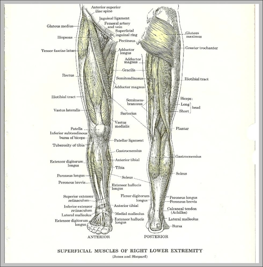
Tag Archives: muscles
Pull Muscles Image
A pulled muscle occurs when a muscle anywhere in the body is stretched beyond its means, leading to slight tearing of the tiny fibers that make up the muscle. A pulled muscle is sometimes known as a strained muscle, and the condition can be quite painful, though some instances of a pulled muscle will result in very little pain.
Pull ups are a compound movement As you can see above, the exercise incorporates multiple muscle groups at one time, opening itself up for the many benefits of compound movements such as: Increased muscle growth – as all of the above muscles are getting a workout at once, rather than isolation exercises targeting one muscle at a time.
What muscles do pull ups work? 1 Lats (latissimus dorsi) 2 Traps (Trapezius) 3 Rhomboids (Rhomboideus major and minor) 4 Posterior deltoid 5 Biceps (Biceps brachii) 6 Teres Major More …
Pull Muscles Image Diagram - Chart - diagrams and charts with labels. This diagram depicts Pull Muscles Image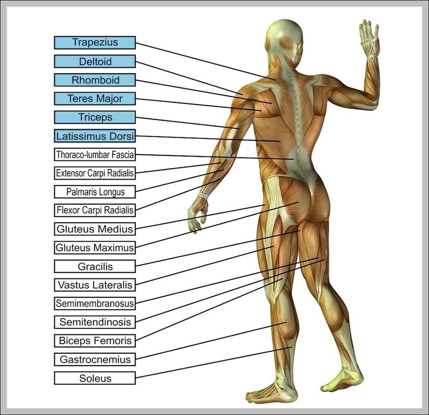
Human Muscles Image
93,451 human body muscles stock photos and images available, or search for anatomy or human anatomy to find more great stock photos and pictures.
Last Updated: Jul 16, 2019 The muscular system is responsible for the movement of the human body. Attached to the bones of the skeletal system are about 700 named muscles that make up roughly half of a person’s body weight. Each of these muscles is a discrete organ constructed of skeletal muscle tissue, blood vessels, tendons, and nerves.
Muscles are the only tissue in the body that has the ability to contract and therefore move the other parts of the body. Related to the function of movement is the muscular system’s second function: the maintenance of posture and body position. Muscles often contract to hold the body still or in a particular position rather than to cause …
Human Muscles Image Diagram - Chart - diagrams and charts with labels. This diagram depicts Human Muscles Image
Arms Muscles Image
27,539 arm muscle anatomy stock photos, vectors, and illustrations are available royalty-free.
What Are the Muscles of Your Arms? There are four main muscles of your arms: biceps, triceps, forearm flexors, and forearm extensors. There are also a handful of other muscles that support these main four.
Anterior arm muscles The body’s anterior muscles tend to be the flexors — they pull your extremities inward, toward your center. So the biceps of the upper arms flex (bend) the elbow, and the forearm flexors on the inside of your forearms flex the wrist and fingers.
Arms Muscles Image Diagram - Chart - diagrams and charts with labels. This diagram depicts Arms Muscles Image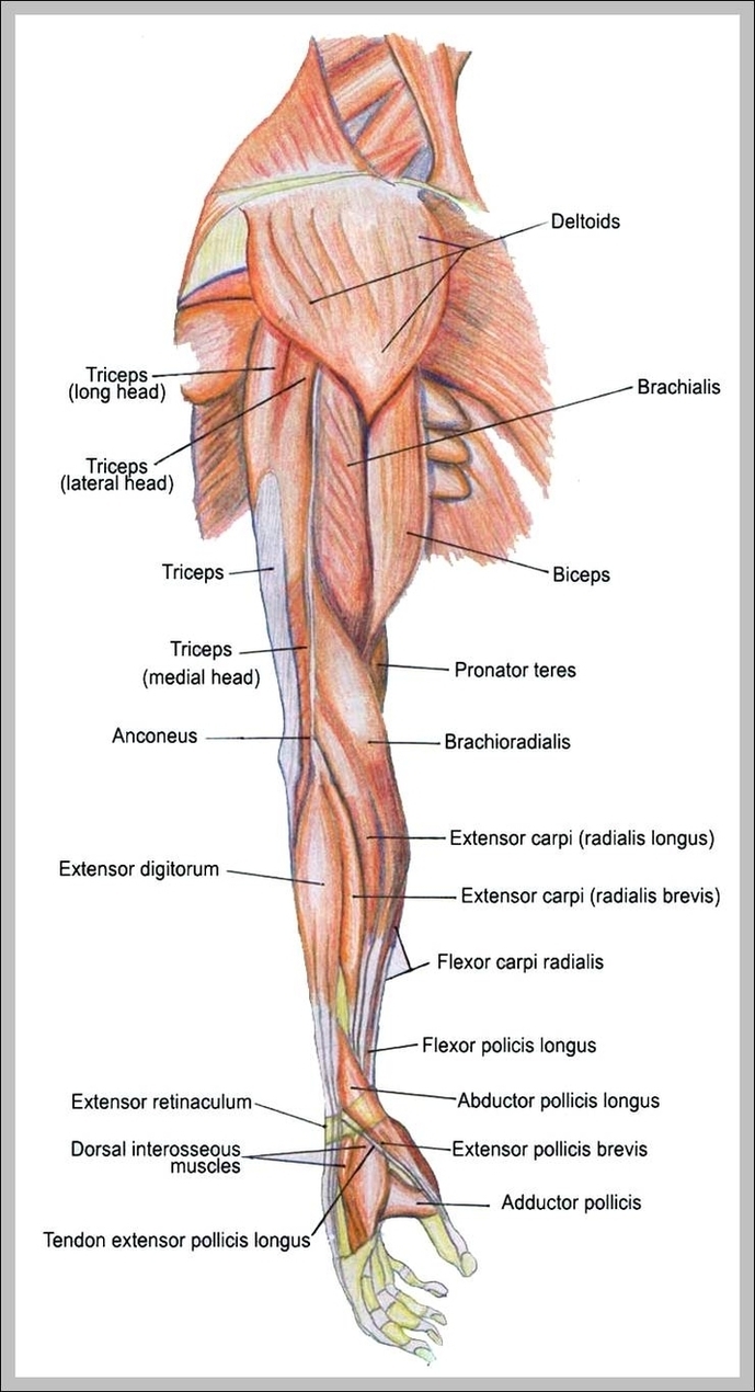
Diagram Of Human Anatomy Muscles Arm Tkdcw Image
The human arm is divided into two main regions, the portion from the elbow to the wrist known as the forearm, and the segment from the shoulder to the elbow referred to as the arm. Arm muscle anatomy enables the arm to perform a variety of movements, including flexion, extension, pronation, and supination.
105,188 human muscle anatomy stock photos, vectors, and illustrations are available royalty-free.
Each of these two sections of the human arm consists of arm muscle anatomy that allows for the flexion, extension, pronation, and supination of the arm, as will be further discussed below. While four muscles are responsible for the upper arm musculature, there are over twenty muscles that move the forearm, wrist, hands, and fingers.
Diagram Of Human Anatomy Muscles Arm Tkdcw Image Diagram - Chart - diagrams and charts with labels. This diagram depicts Diagram Of Human Anatomy Muscles Arm Tkdcw Image
Human Anatomy Muscles Arm Polycount Madell Forum Image
Human Anatomy Muscles Arm Polycount Madell Forum Image Diagram - Chart - diagrams and charts with labels. This diagram depicts Human Anatomy Muscles Arm Polycount Madell Forum Image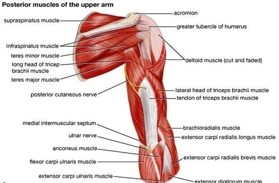
Picture Of Shoulder Muscles Image
124,600 shoulder muscles stock photos, vectors, and illustrations are available royalty-free. See shoulder muscles stock video clips
For that reason, and because of the dexterity of the shoulder joint itself, the musculature of the shoulder is complex, ranging from massive prime mover muscles to finer stabilizer and fixator muscles. The shoulder (or humeroscapular) joint is formed by the articulation of the head of the humerus with the scapula.
On the anterior side of the shoulder, the coracobrachialis, serratus anterior, pectoralis major, and pectoralis minor muscles work as a group to flex and adduct the scapula and humerus anteriorly toward the sternum. The latissimus dorsi and teres major on the posterior side extend and adduct the arm towards the vertebrae of the back.
Picture Of Shoulder Muscles Image Diagram - Chart - diagrams and charts with labels. This diagram depicts Picture Of Shoulder Muscles Image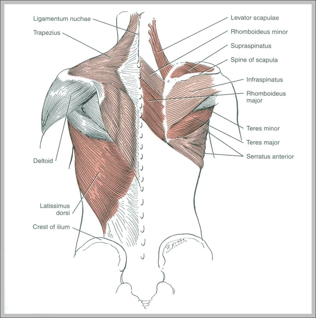
Muscles Pictures Image
226,053 human muscle stock photos and images available, or search for human muscle anatomy or human muscle tissue to find more great stock photos and pictures. Man with extended arm. Illustrated representation of the structure and musculature of the human arm. Studio man portrait. human muscle stock pictures, royalty-free photos & images
Forearms – Anatomy Muscles Rhomboid minor and rhomboid major, levator scapulae and latissimus dorsi muscles – didactic board of anatomy of human bony and muscular system, posterior view Abs – Anatomy Muscles Leg muscles of the man Human Body Organs (Lungs) 3D Triceps – Anatomy Muscles Hand muscle connection with brain
Vicky Timón, a yoga expert and author of âEncyclopedia of Pilates Exercises,â created 36 beautiful illustrations to show you which muscles are affected by different stretches. Camel Pose: Best reserved for those with good flexibility, the muscles highlighted in this stretch are the rectus abdominus and the external obliques.
Muscles Pictures Image Diagram - Chart - diagrams and charts with labels. This diagram depicts Muscles Pictures Image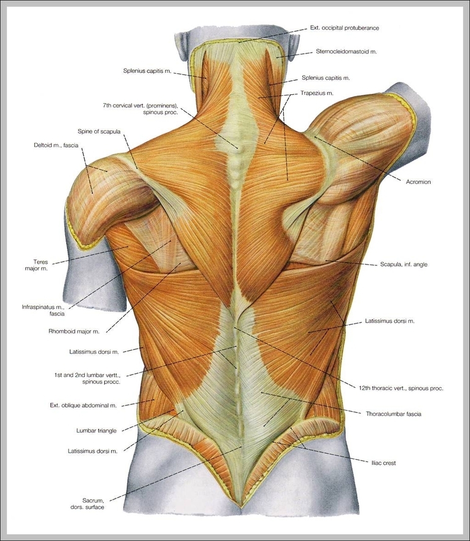
Upper Extremity Muscles Diagram Image
Upper limb muscles and movements. The upper limb (upper extremity) is truly a complex part of human anatomy. It is best studied broken down into its components: regions, joints, muscles, nerves, and blood vessels.
Upper limb muscles and movements. 1 Scapular region. The scapular region lies on the posterior surface of the thoracic wall. It may seem strange that it is included in the anatomy of the … 2 Shoulder. 3 Arm (brachium) 4 Forearm flexors. 5 Forearm extensors. More items
The upper arm is located between the shoulder joint and elbow joint. It contains four muscles – three in the anterior compartment (biceps brachii, brachialis, coracobrachialis), and one in the posterior compartment (triceps brachii). In this article, we shall look at the anatomy of the muscles…
Upper Extremity Muscles Diagram Image Diagram - Chart - diagrams and charts with labels. This diagram depicts Upper Extremity Muscles Diagram Image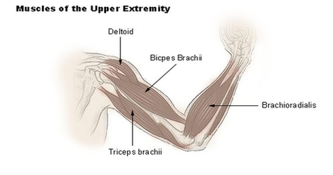
Muscles Of The Human Body Diagram Image
105,188 human muscle anatomy stock photos, vectors, and illustrations are available royalty-free.
Last Updated: Jul 16, 2019 The muscular system is responsible for the movement of the human body. Attached to the bones of the skeletal system are about 700 named muscles that make up roughly half of a person’s body weight. Each of these muscles is a discrete organ constructed of skeletal muscle tissue, blood vessels, tendons, and nerves.
Tibialis Anterior Muscle Tibialis Posterior Muscle Vastus Lateralis Muscle Vastus Medialis Muscle GENERAL Cross-Sections of Muscles of the Arm and Leg Muscle Cell Types Pronation/Supination Push/Pull Muscles Change Current View Angle Muscular System (Male View) Muscular System (Male Posterior View) Muscular System (Posterior View)
Muscles Of The Human Body Diagram Image Diagram - Chart - diagrams and charts with labels. This diagram depicts Muscles Of The Human Body Diagram Image
Human Anatomy Muscles Image
Human Anatomy Muscles Image Diagram - Chart - diagrams and charts with labels. This diagram depicts Human Anatomy Muscles Image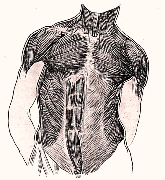
Shoulder Anatomy Muscles Diagram Image
shoulder joint anatomy shoulder joint anatomy. Bones (Scapula, Humerus, Coracoid process, Acromion , Muscle (Biceps, Supraspinatus) and ligament (Coracohumeral) shoulder anatomy stock illustrations shoulder joint anatomy.
13,987 shoulder anatomy stock photos and images available, or search for shoulder anatomy muscles to find more great stock photos and pictures. Man with extended arm. Illustrated representation of the structure and musculature of the human arm. Studio man portrait. shoulder anatomy stock pictures, royalty-free photos & images
Toggle Anatomy System. Surrounding the rotator cuff muscles are many groups of muscles that work together to produce the various movements of the shoulder. Located superior to the shoulder joint, the deltoid muscle works with the supraspinatus to abduct the arm at the shoulder. On the anterior side of the shoulder, the coracobrachialis,…
Shoulder Anatomy Muscles Diagram Image Diagram - Chart - diagrams and charts with labels. This diagram depicts Shoulder Anatomy Muscles Diagram Image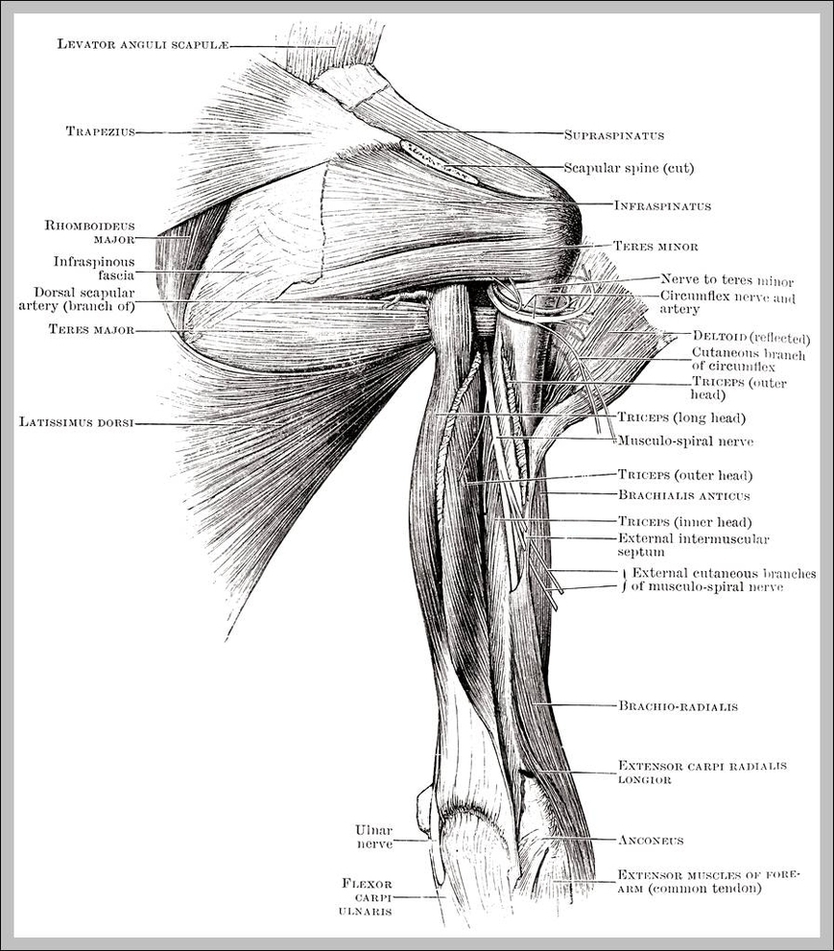
Pictures Of Skeletal Muscles Image
292 skeletal muscle stock photos and images available, or search for skeletal muscle fibers or skeletal muscle fiber to find more great stock photos and pictures. Fascicle Muscle.
Fascicle Muscle. Representation Of A Fascicle Muscle A Muscle Is Constituted Of Various Fascicle Muscles Constituted Of Various Fiber Muscles Largely… Muscle Spindle, Neuromuscular Spindle And Proprioception Is The Perception Of The Position Of The Different Parts Of The Body In Space, It Is…
An example of an anatomical and physical movement process where the biceps are contracted and the triceps are relaxed, the biceps is relaxed and the triceps are contracted. Skeletal Muscle stock illustrations An example of an anatomical and physical movement process where…
Pictures Of Skeletal Muscles Image Diagram - Chart - diagrams and charts with labels. This diagram depicts Pictures Of Skeletal Muscles Image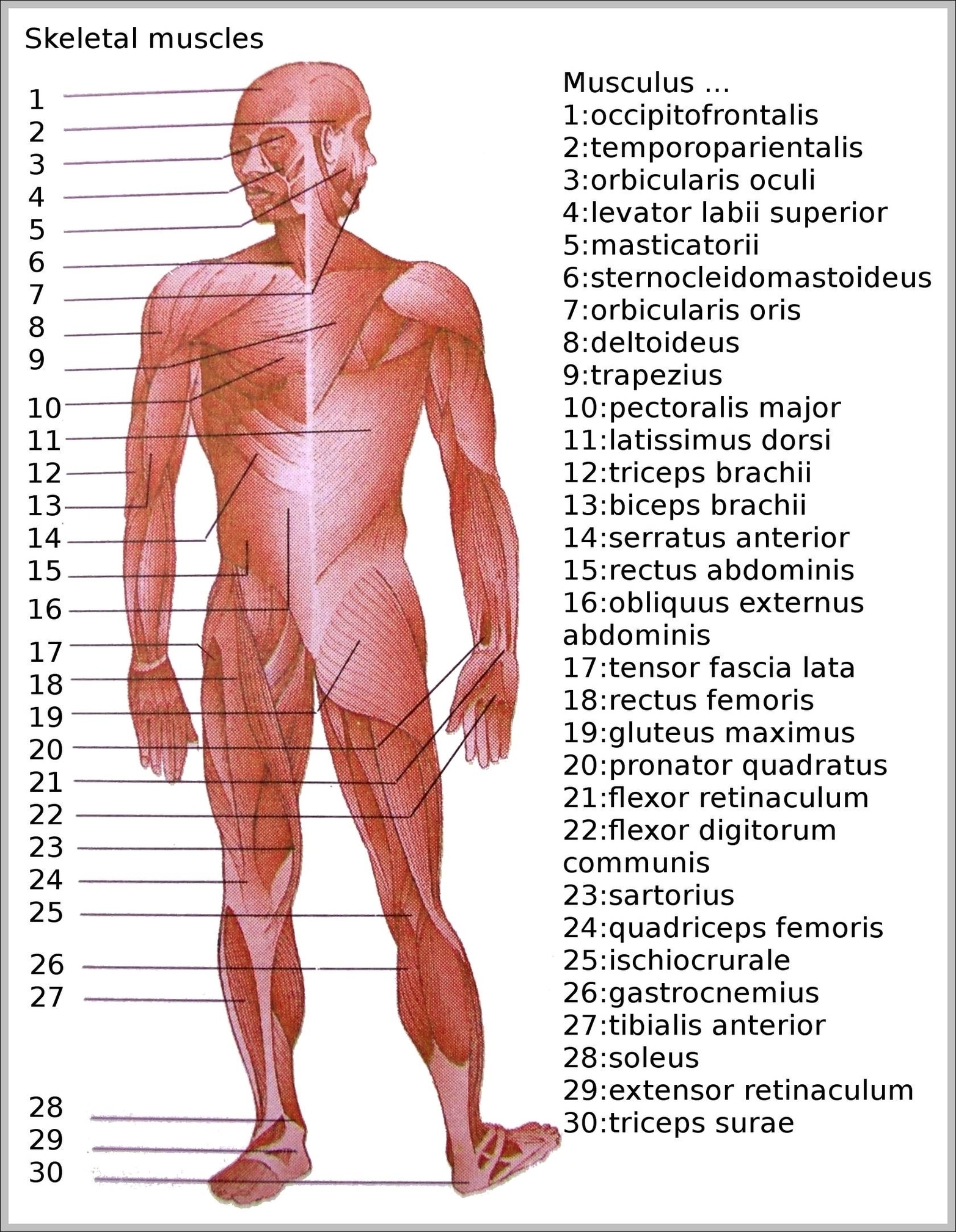
Diagram Human Eye Muscles Image
The Human Eye (Eyeball) Diagram, Parts and Pictures. The human eye consists of the eyeball, optic nerve, orbit and appendages (eyelids, extraocular muscles and lacrimal glands). While the eyeball is the actual sensory organ, the other parts of of the eye are equally important in maintaining the health and function of the eye as a whole.
A series of muscles helps the eye move. The first set is the superior and inferior rectus muscles, which allow upward and downward motion. The medial and lateral rectus muscles allow the eye to move from side to side while staying level. The superior and inferior oblique muscles let it move up or down and to the side.
Structure Of Eye. The eye is one of the important sensory organs in the human body. It is mainly responsible for vision, differentiation of color (the human eye can differentiate approximately 10 – 12 million colors) and maintaining the biological clock of the human body. The human eye can be compared to a camera as both functions by gathering,…
Diagram Human Eye Muscles Image Diagram - Chart - diagrams and charts with labels. This diagram depicts Diagram Human Eye Muscles Image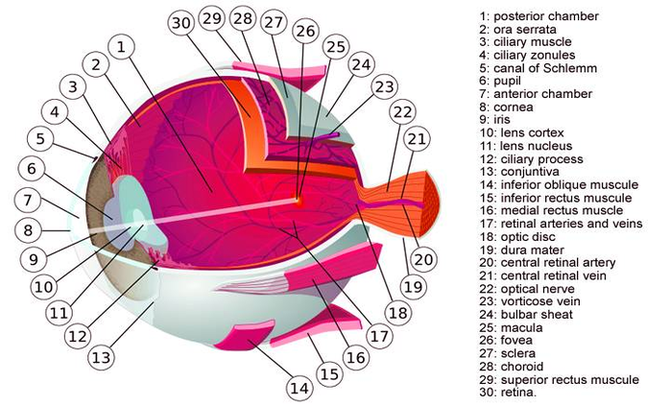
Pictures Of Body Muscles Image
93,451 human body muscles stock photos and images available, or search for anatomy or human anatomy to find more great stock photos and pictures.
Forearms – Anatomy Muscles Rhomboid minor and rhomboid major, levator scapulae and latissimus dorsi muscles – didactic board of anatomy of human bony and muscular system, posterior view Abs – Anatomy Muscles Leg muscles of the man Human Body Organs (Lungs) 3D Triceps – Anatomy Muscles Hand muscle connection with brain
Plastic model a human body, clear with inside system on one side, and muscle and flesh on the other seen from back Human body. Plastic model of a human body, clear with inside system on one side, and muscle and flesh on the other Human body.
Pictures Of Body Muscles Image Diagram - Chart - diagrams and charts with labels. This diagram depicts Pictures Of Body Muscles Image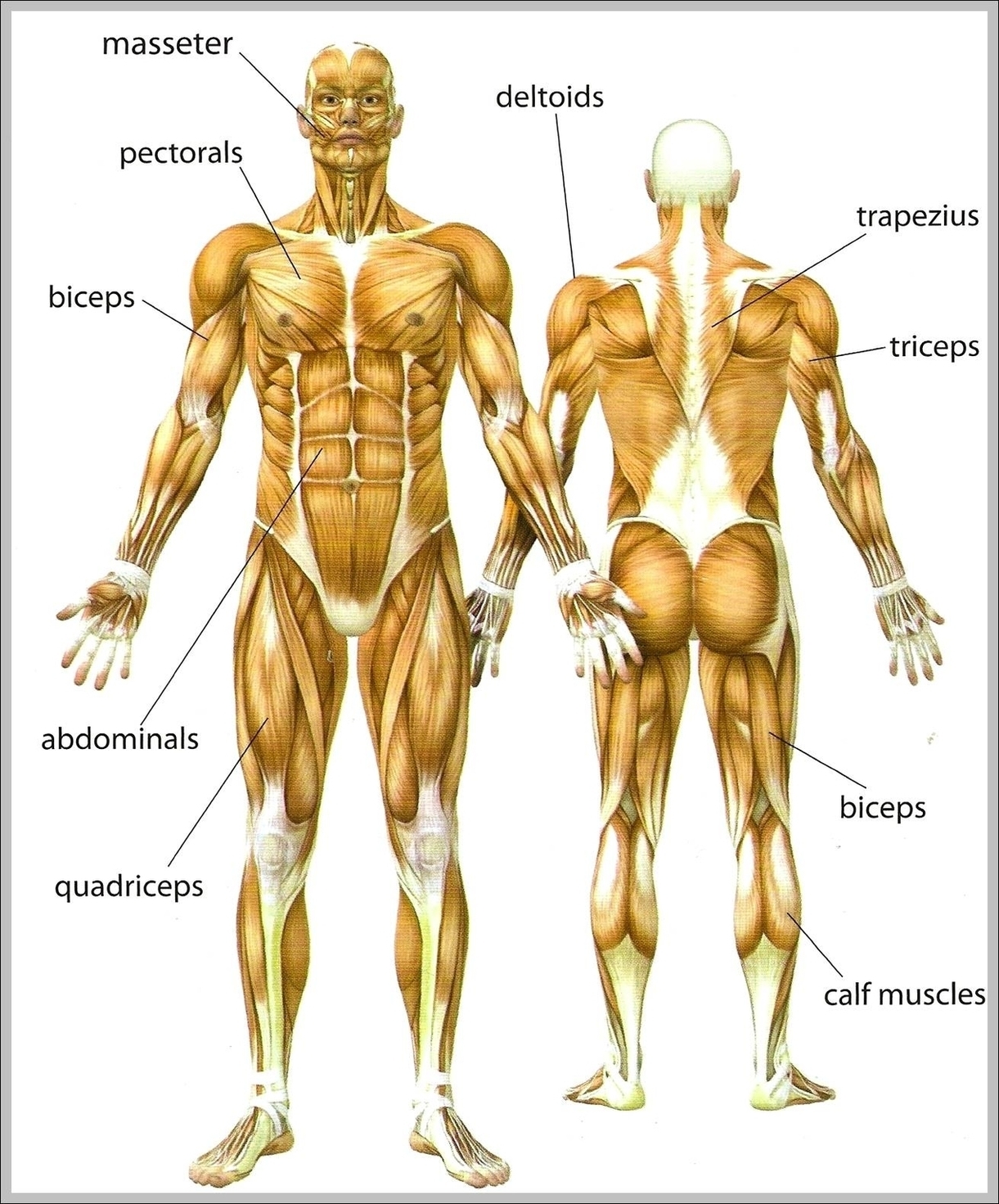
Anatomy Arm Muscles Image
Anatomy Arm Muscles Image Diagram - Chart - diagrams and charts with labels. This diagram depicts Anatomy Arm Muscles Image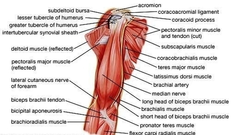
Muscles Stability Image
Following are the five major muscles / muscle groups of sacroiliac stabilization that should be assessed and likely worked with manual therapy when the client presents with a sacroiliac joint condition. Piriformis Gluteus Maximus (superior deep fibers) Coccygeus and Levator Ani Paraspinals Hamstrings
Stabilizer muscles do just what their name implies in that they help stabilize joints and parts of the body so that movement patterns are efficient in a kinematic sense,” says John Mikula, CSCS and a consultant through Tactical Speed and Strength.
The hip joint is one of the most flexible joints in the entire human body. The many muscles of the hip provide movement, strength, and stability to the hip joint and the bones of the hip and thigh. These muscles can be grouped based upon their location and function. The four groups are the anterior group, the posterior group,…
Muscles Stability Image Diagram - Chart - diagrams and charts with labels. This diagram depicts Muscles Stability Image
Muscles In Back Diagram Image
25,649 back muscle anatomy stock photos, vectors, and illustrations are available royalty-free.
There are three different muscle groups found in the back: the superficial group, the deep group, and the intermediate group. Muscles found in the superficial group include rhomboid major, rhomboid minor, levator scapulae, trapezius, latissimus dorsi.
The back supports the weight of the body, allowing for flexible movement while protecting vital organs and nerve structures. This article looks at the anatomy of the back, including bones, muscles, and nerves. It also covers some common conditions and injuries that can affect the back.
Muscles In Back Diagram Image Diagram - Chart - diagrams and charts with labels. This diagram depicts Muscles In Back Diagram Image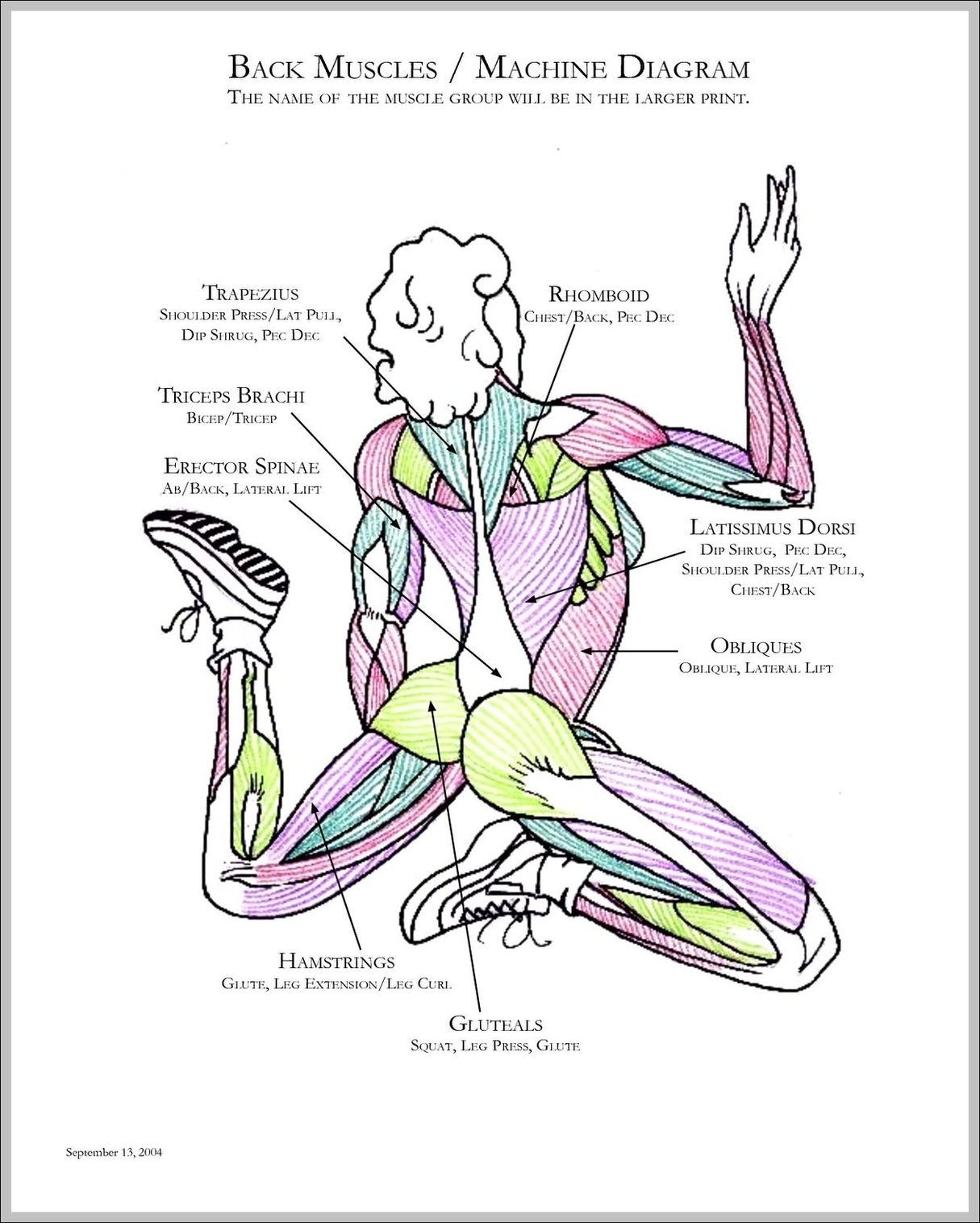
Anatomy Of The Leg Muscles Image
72,510 leg anatomy stock photos, vectors, and illustrations are available royalty-free.
Leg muscles are bundles of fibrous tissue that contract and relax to exert forces on bones and move the legs The main muscle groups in the legs are: quadriceps, hamstrings, adductors in the upper leg or thigh, and the calves in the lower legs.
The legs are the lower extremities that allow for standing, walking, running, and more. Their movement is produced by the contraction and relaxation of leg muscles and their connection to the skeleton through tendons. The movement of the legs produced by these muscles happens around the leg joints, particularly the hips, knees and ankles.
Anatomy Of The Leg Muscles Image Diagram - Chart - diagrams and charts with labels. This diagram depicts Anatomy Of The Leg Muscles Image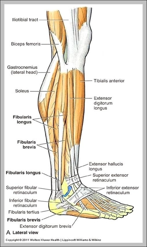
Muscles Diagram Image
Last Updated: Jul 16, 2019 The muscular system is responsible for the movement of the human body. Attached to the bones of the skeletal system are about 700 named muscles that make up roughly half of a person’s body weight. Each of these muscles is a discrete organ constructed of skeletal muscle tissue, blood vessels, tendons, and nerves.
Tibialis Anterior Muscle Tibialis Posterior Muscle Vastus Lateralis Muscle Vastus Medialis Muscle GENERAL Cross-Sections of Muscles of the Arm and Leg Muscle Cell Types Pronation/Supination Push/Pull Muscles Change Current View Angle Muscular System (Male View) Muscular System (Male Posterior View) Muscular System (Posterior View)
Finally, the direction in which the muscle fibers run can be used to identify a muscle. In the abdominal region, there are several sets of wide, flat muscles.
Muscles Diagram Image Diagram - Chart - diagrams and charts with labels. This diagram depicts Muscles Diagram Image