Metastatic Pancreatic Neuroendocrine Image

What Is a Pancreatic Neuroendocrine Tumor? Pancreatic neuroendocrine tumors (NETs), or islet cell tumors, are a type of cancer that starts in the pancreas. (Cancer starts when cells in the body begin to grow out of control. To learn more about how cancers start and spread, see What Is Cancer?)

The metastatic tumor is the same type of tumor as the primary tumor. For example, if a pancreatic neuroendocrine tumor spreads to the liver, the tumor cells in the liver are actually neuroendocrine tumor cells. The disease is metastatic pancreatic neuroendocrine tumor, not liver cancer. If playback doesn’t begin shortly, try restarting your device.

Tumor grade. Pancreatic neuroendocrine tumors (NETs) are classified by tumor grade, which describes how quickly the cancer is likely to grow and spread. Grade 1 (also called low-grade or well-differentiated) neuroendocrine tumors have cells that look more like normal cells and are not multiplying quickly.

Metastatic Pancreatic Neuroendocrine Image Diagram - Chart - diagrams and charts with labels. This diagram depicts Metastatic Pancreatic Neuroendocrine Image

Metastatic Pancreatic Neuroendocrine Image

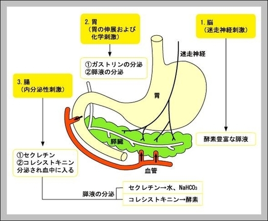
Pancreatic Juice Image

Pancreatic juice is a liquid secreted by the pancreas, which contains a variety of enzymes, including trypsinogen, chymotrypsinogen, elastase, carboxypeptidase, pancreatic lipase, nucleases and amylase.

Pancreatic juice. Pancreatic juice is secreted into the duodenum through duodenal papillae. Some individuals have also an accessory duct, named accessory pancreatic duct, which may be functional (that is, it also empties the contents of the exocine pancreas into the duodenum) or non-functional.

Pancreatic juice is an exceptionally rich source of proteins released from pancreatic cancer cells and therefore an ideal specimen for biomarkers discovery [4].

Pancreatic Juice Image Diagram - Chart - diagrams and charts with labels. This diagram depicts Pancreatic Juice Image

Pancreatic Juice Image

Pancreatic System Image

Anatomy of the pancreas; drawing shows the pancreas, stomach, spleen, liver, gallbladder, bile ducts, colon, and small intestine. An inset shows the head, body, and tail of the pancreas. The bile duct and pancreatic duct are also shown. Anatomy of the pancreas. The pancreas has three areas: head, body, and tail.

1,908 pancreas anatomy stock photos and images available, or search for kidney anatomy or liver anatomy to find more great stock photos and pictures.

Pancreatitis: The pancreas becomes inflamed and damaged by its own digestive chemicals. Swelling and death of tissue of the pancreas can result. Although alcohol or gallstones can contribute, sometimes a cause for pancreatitis is never found. Pancreatic pseudocyst: After a bout of pancreatitis, a fluid-filled cavity called a pseudocyst can form.

Pancreatic System Image Diagram - Chart - diagrams and charts with labels. This diagram depicts Pancreatic System Image

Pancreatic System Image