X Advanced Heart Development Timeline Ga Image

This human anatomy diagram with labels depicts and explains the details and or parts of the X Advanced Heart Development Timeline Ga Image. Human anatomy diagrams and charts show internal organs, body systems, cells, conditions, sickness and symptoms information and/or tips to ensure one lives in good health.

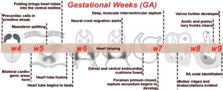
Early development. The heart derives from embryonic mesodermal germ-layer cells that differentiate after gastrulation into mesothelium, endothelium, and myocardium. Mesothelial pericardium forms the outer lining of the heart. The inner lining of the heart, lymphatic and blood vessels, develop from endothelium.

Development of the Cardiovascular System 1 Primitive Heart Tube. The development of the heart begins with the formation of the primitive heart tube following the folding of the embryo during the end of the third week. 2 Atria. … 3 Aortic Arches. … 4 Septation of the Heart. … 5 Circulatory Shunts. …

Heart development. This begins with the formation of two endocardial tubes which merge to form the tubular heart, also called the primitive heart tube, that loops and septates into the four chambers and paired arterial trunks that form the adult heart. The heart is the first functional organ in vertebrate embryos, and in the human,…

X Advanced Heart Development Timeline Ga Image

X Advanced Heart Development Timeline Ga Image


Tags: , , , , ,