Pleural Cavity Image

This human anatomy diagram with labels depicts and explains the details and or parts of the Pleural Cavity Image. Human anatomy diagrams and charts show internal organs, body systems, cells, conditions, sickness and symptoms information and/or tips to ensure one lives in good health.

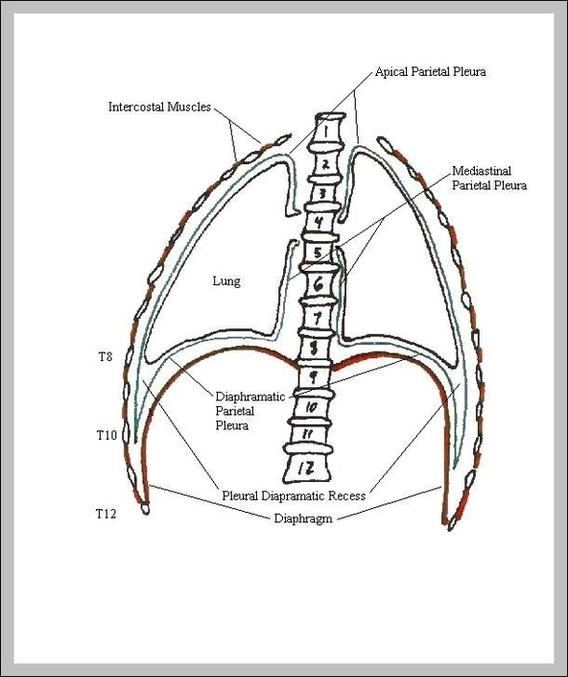
The pleural cavity is bounded by a double layered serous membrane called pleura. Pleura is formed by an inner visceral pleura and an outer parietal layer. Between these two membranous layers is a small amount of serous fluid held within the pleural cavity. This lubricated cavity allows the lungs to move freely during breathing.

Pleural space The pleural cavity surrounds the lungs in the thoracic cavity. There are two pleural cavities, one for each lung on the right and left sides of the mediastinum. Each pleural cavity and it’s enclosed lung are lined by a serous membrane called pleura.

The pleurae are two layers of serous membrane that form the boundaries of the pleural cavity. There are two types of pleura; parietal and visceral.

Pleural Cavity Image

Pleural Cavity Image


Tags: ,