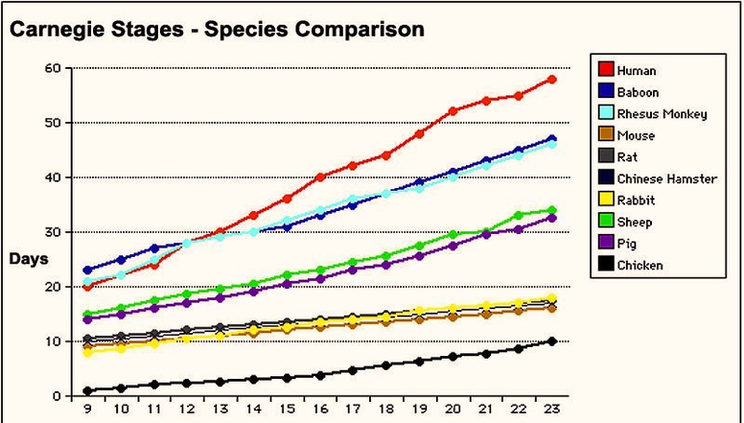
About Translations ) Carnegie stages are named after the famous US Institute which began collecting and classifying embryos in the early 1900’s. Stages are based on the external and/or internal morphological development of the embryo, and are not directly dependent on either age or size.
There are only two stage 3 embryos in the Carnegie collection. There are four characteristic processes that CS3 embryos go through cavitation, collapse and expansion, hatching, and discarding of cells. The initiation of cavitation indicates the start of CS3.
Carnegie stage 1 is the unicellular embryo. This stage is divided into three substages. Primordial embryo. All the genetic material necessary for a new individual, along with some redundant chromosomes, are present within a single plasmalemma. Penetration of the fertilising sperm allows the oocyte to resume meiosis and the polar body is extruded.
Carnegie Stages Species Comparison Image

