Trimester Image

This human anatomy diagram with labels depicts and explains the details and or parts of the Trimester Image. Human anatomy diagrams and charts show internal organs, body systems, cells, conditions, sickness and symptoms information and/or tips to ensure one lives in good health.

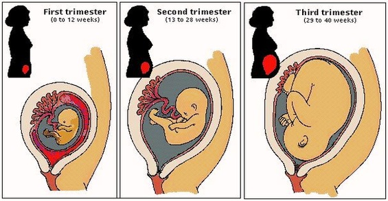
Pregnancy has three trimesters, each of which is marked by specific fetal developments. A pregnancy is considered full-term at 40 weeks; infants delivered before the end of week 37 are considered premature. Premature infants may have problems with their growth and development, as well as difficulties in breathing and digesting.

Fetal Development Milestones: Head growing larger, and structures that will form the brain can be identified. Nostrils and lenses of the eyes develop. What You’re Seeing: During this week of the first trimester, you can see baby-to-be is developing in a bubble within the gestational sac.

(1st, 2nd, and 3rd Trimester) Conception to about the 12th week of pregnancy marks the first trimester. The second trimester is weeks 13 to 27, and the third trimester starts about 28 weeks and lasts until birth. This slide show will discuss what occurs to both the mother and baby during each trimester.

Trimester Image

Trimester Image


Tags: