Thoracic Vertebra

This human anatomy diagram with labels depicts and explains the details and or parts of the Thoracic Vertebra. Human anatomy diagrams and charts show internal organs, body systems, cells, conditions, sickness and symptoms information and/or tips to ensure one lives in good health.

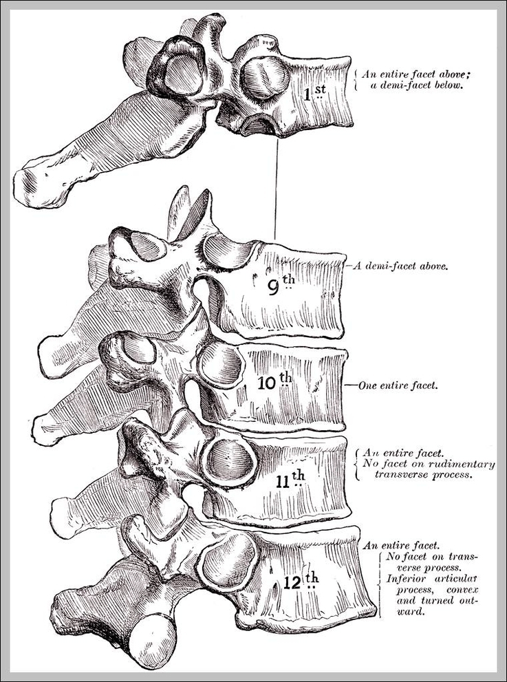
In vertebrates, thoracic vertebrae compose the middle segment of the vertebral column, between the cervical vertebrae and the lumbar vertebrae.

The thoracic spine has 12 vertebrae stacked on top of each other, labeled from T1 down to T12. These vertebrae form the foundation of the thoracic region’s sturdy spinal column that supports the neck above, the rib cage, soft tissues, flexible joints, blood vessels, and nerves.

April 19, 2019. The thoracic vertebrae are bones located between the cervical and lumbar vertebrae. There are 12 thoracic vertebrae in humans, and these bones increase in size as you move down the body.

Thoracic Vertebra

thoracic vertebra