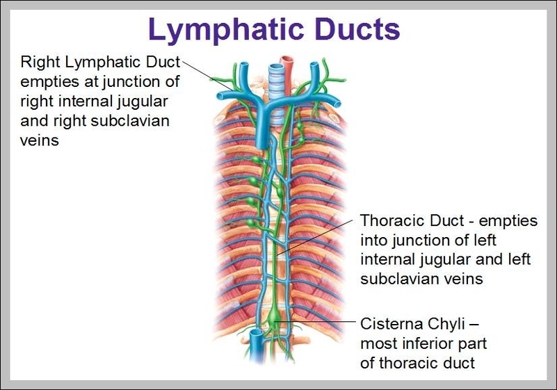
The action of breathing helps chyle flow up the thoracic duct. The duct also contains smooth muscle within its walls, as well as interval valves (much like large veins), which prevent backflow of lymph. Need to refresh your knowledge of the basic anatomy of the digestive system? Our digestive system quizzes and free learning tools have your back.
Anatomical terminology. In human anatomy, the thoracic duct is the larger of the two lymph ducts of the lymphatic system. It is also known as the left lymphatic duct, alimentary duct, chyliferous duct, and Van Hoorne’s canal.
It is also thin and easily torn. The thoracic duct drains lymph from the right and left descending thoracic lymph trunks, originating from the lower 6 intercostal spaces (6 to 11). The duct also receives lymph from intercostal spaces 1 to 5 via the upper intercostal lymph trunks. Additional tributaries include the:
Thoracic Duct Function

