Cerebral Peduncle Image

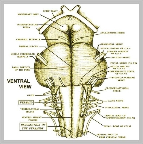
The cerebral peduncle is made of a mass of nerve fibers, and there is one peduncle on each side of the brain. The term ‘cerebral’ means it is related to the brain.

The brain peduncles they are brain casts made up entirely of nerves. Each human brain has two cerebral peduncles that are joined by an interpeduncular fossa. The cerebral peduncles are located in the upper region of the brainstem, just above the annular pons.

Cerebral penduncle. Many other nerve bundles also connect to the pons. Cerebral peduncles help transport nerve impulses from the higher part of the brain (cortex) and the brain stem, or lower part of the brain, to other areas of the central nervous system.

Cerebral Peduncle Image Diagram - Chart - diagrams and charts with labels. This diagram depicts Cerebral Peduncle Image

Cerebral Peduncle Image

Cerebral Peduncles Image

The Cerebral peduncles Are brain cylinders composed entirely of nerves. Each human brain has two cerebral peduncles that are united by an interpeduncular fossa. The cerebral peduncles are located in the superior region of the brainstem, just above the annular protuberance.

Cerebral penduncle. Many other nerve bundles also connect to the pons. Cerebral peduncles help transport nerve impulses from the higher part of the brain (cortex) and the brain stem, or lower part of the brain, to other areas of the central nervous system.

Cerebral penduncle. The peduncles adjust the commands by taking into account where the body parts currently are located before directing the movement, and they sometimes slow down the movement. When there is an injury to cerebral peduncles, the symptoms of the injury show up in the part of the body related to the injured peduncle.

Cerebral Peduncles Image Diagram - Chart - diagrams and charts with labels. This diagram depicts Cerebral Peduncles Image

Cerebral Peduncles Image

The Cerebral Cortex Image

1,246 cerebral cortex stock photos and images available, or search for cerebral cortex neuron to find more great stock photos and pictures. Normal Cerebral Cortex. Computer Generated 3D Image. Two Fissures Of Rolando Are Visible. Department Of Morphological And Functional Imaging, Led By…

The cerebral cortex (cortex of the brain) is the outer grey matter layer that completely covers the surface of the two cerebral hemispheres. It is about 2 to 4 mm thick and contains an aggregation of nerve cell bodies. This layer is thrown into complex folds, with elevations called gyri  and grooves known as sulci.

The cerebral cortex is folded in a way that allows a large surface area of neural tissue to fit within the confines of the neurocranium. When unfolded in the human, each hemispheric cortex has a total surface area of about 1.3 square feet (0.12 m 2 ). The folding is inward away from the surface of the brain,…

The Cerebral Cortex Image Diagram - Chart - diagrams and charts with labels. This diagram depicts The Cerebral Cortex Image

The Cerebral Cortex Image