Thoracic Cage Diagram Image

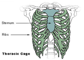
The angles of the ribs form the most posterior extent of the thoracic cage. In the anatomical position, the angles align with the medial border of the scapula. A shallow costal groove for the passage of blood vessels and a nerve is found along the inferior margin of each rib. The bony ribs do not extend anteriorly completely around to the sternum.

The thoracic cage (rib cage) forms the thorax (chest) portion of the body. It consists of the 12 pairs of ribs with their costal cartilages and the sternum ( Figure 7.5.1 ).

The ribs are anchored posteriorly to the 12 thoracic vertebrae (T1–T12). The thoracic cage protects the heart and lungs. Figure 7.5.1 – Thoracic Cage: The thoracic cage is formed by the (a) sternum and (b) 12 pairs of ribs with their costal cartilages.

Thoracic Cage Diagram Image Diagram - Chart - diagrams and charts with labels. This diagram depicts Thoracic Cage Diagram Image

Thoracic Cage Diagram Image