Posterior Commissure Image

This human anatomy diagram with labels depicts and explains the details and or parts of the Posterior Commissure Image. Human anatomy diagrams and charts show internal organs, body systems, cells, conditions, sickness and symptoms information and/or tips to ensure one lives in good health.

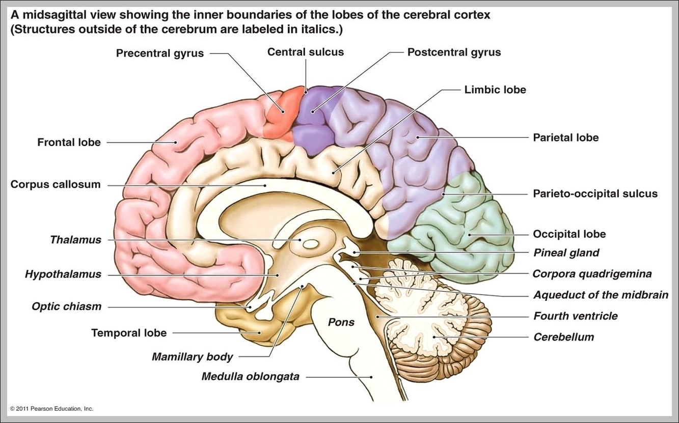
Anatomical terms of neuroanatomy. [edit on Wikidata] The posterior commissure (also known as the epithalamic commissure) is a rounded band of white fibers crossing the middle line on the dorsal aspect of the rostral end of the cerebral aqueduct. It is important in the bilateral pupillary light reflex.

Posteriorly the labia are not really joined, but appear to become lost in the neighboring integument, ending close to, and nearly parallel with, each other. Together with the connecting skin between them, they form the posterior labial commissure or posterior boundary of the pudendum. There is no image containing this anatomical part yet.

On the basis of clinical observations, the nucleus of the posterior commissure (PCom) has long been suspected as being involved in the generation of upward eye movements, since lesions in patients resulted in upward gaze paralysis ( Christoff, 1974 ). To date the role of the PCom in vertical gaze has not been fully explained.

Posterior Commissure Image

Posterior Commissure Image


Tags: ,