Picture Of a Spine

This human anatomy diagram with labels depicts and explains the details and or parts of the Picture Of a Spine. Human anatomy diagrams and charts show internal organs, body systems, cells, conditions, sickness and symptoms information and/or tips to ensure one lives in good health.

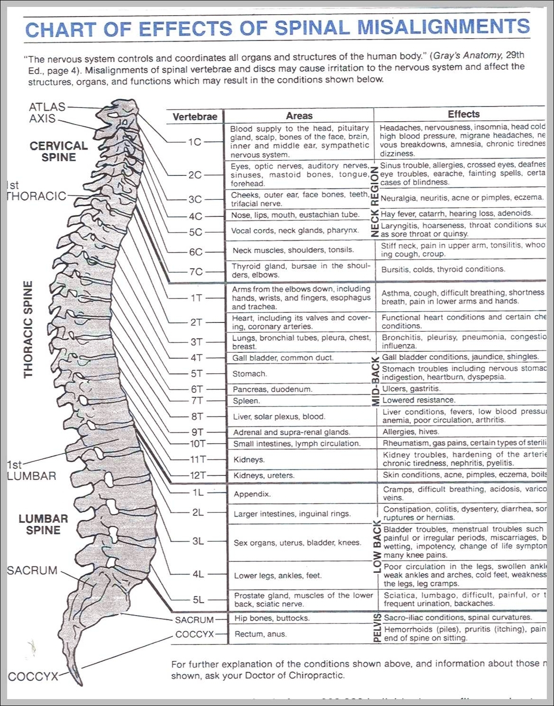
First are the vertebrae of the spine, and underneath those are three layers of tough membrane called the meninges. The meninges surround both brain and spinal cord and are filled with a liquid called cerebrospinal fluid.

Last Updated: Jul 3, 2018. Stretching down the midline of the trunk from the base of the skull to the coccyx, the spine plays an extremely important role in our bodies as it supports the upper body’s weight; provides posture while allowing for movement and flexibility; and protects the spinal cord.

Your spine has spaces in it for your spinal cord and the nerves that branch out from it. When those spaces shrink, bones can press against nerves. You might not even notice it, but any time nerves are messed with, you could have pain, tingling, or numbness, or your muscles might seem weak. Osteoarthritis is the most common cause of spinal stenosis.

Picture Of a Spine

picture of a spine