Hallux Valgus Ray Image

This human anatomy diagram with labels depicts and explains the details and or parts of the Hallux Valgus Ray Image. Human anatomy diagrams and charts show internal organs, body systems, cells, conditions, sickness and symptoms information and/or tips to ensure one lives in good health.

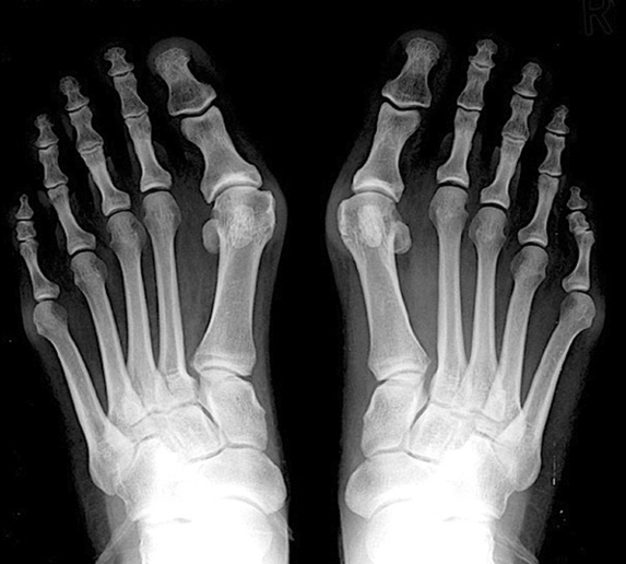
Hallux Valgus, commonly referred to as a bunion, is a complex valgus deformity of the first ray that can cause medial big toe pain and difficulty with shoe wear. Diagnosis is made clinically with presence of a hallux that rests in a valgus and pronated position.

A clinical image is shown in Figure A and a radiograph is shown in Figure B. The hallux valgus angle (HVA) is measured at 23 degrees and the intermetatarsal angle (IMA) is measured at 12 degrees. Which of the following surgical interventions is most appropriate for correction of her deformities?

The combined deformities are present to some degree in most patients. Hallux valgus may be a result of metatarsus primus varus, and surgical correction is often based on this theory. Non-surgical treatment involves the use of accommodative footwear.

Hallux Valgus Ray Image

Hallux Valgus Ray Image


Tags: , ,