External Iliac Image

This human anatomy diagram with labels depicts and explains the details and or parts of the External Iliac Image. Human anatomy diagrams and charts show internal organs, body systems, cells, conditions, sickness and symptoms information and/or tips to ensure one lives in good health.

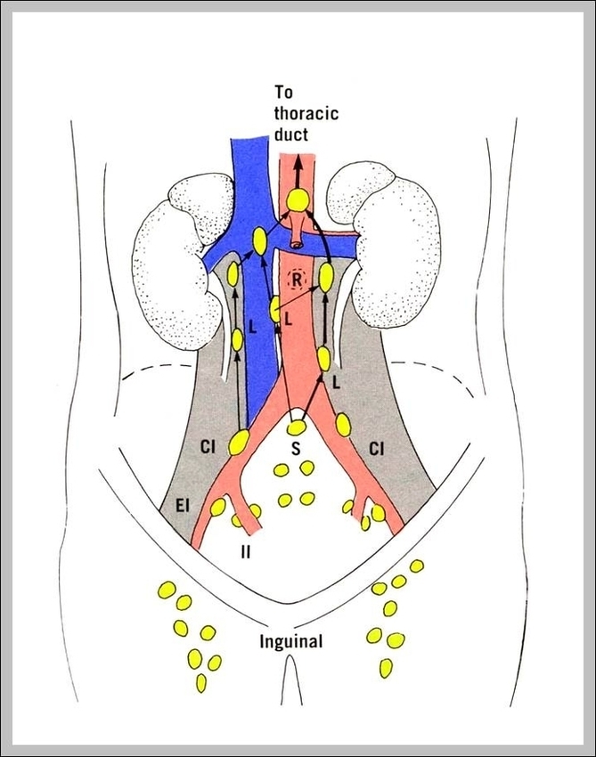
External iliac vessels and their branches in a cadaver: The external iliac artery originates from the common iliac artery. Arteries have thick, muscular walls, hence they have a firmer consistency compared to veins. The inferior epigastric artery branches from the external iliac artery just posterior to the inguinal ligament.

The external iliac lymph nodes can be found surrounding the external iliac artery and act as the draining nodes for several regions of the pelvis and lower limb.

In the abdomen, the aorta splits into the right and left common iliac arteries. At the pelvic brim, each common iliac artery splits into the internal and external iliac arteries. Each external iliac artery courses downward and laterally, turning into the femoral artery, which supplies each leg.

External Iliac Image

External Iliac Image


Tags: ,