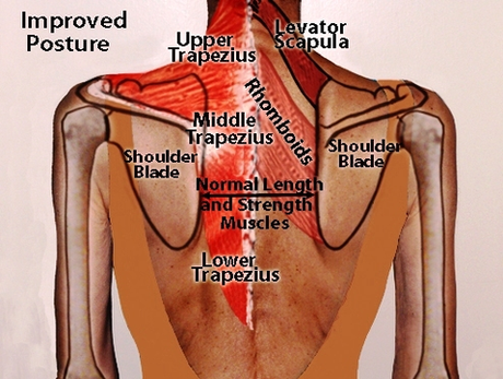
The back consists of the spine, spinal cord, muscles, ligaments, and nerves. These structures work together to support the body, enable a range of movements, and send messages from the brain to the rest of the body. Conditions or injuries affecting the back can range from mild to severe. Back Posture Diagram Image Diagram - Chart - diagrams and charts with labels. This diagram depicts Back Posture Diagram Image and explains the details of Back Posture Diagram Image.
Back Posture Diagram Image