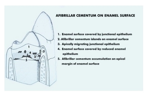
Afibrillar cementum By the origin of the matrix fibers Extrinsic fiber (from PDL fibroblasts) Intrinsic fiber (from cementoblasts) Mixed fiber (contains both of the above) Examples: Acellular, afibrillar cementum(coronal cementum of human teeth) Mostly composed of mineralized ground substance. Free of cells or fibers.
In the cemento-enamel junction, the cementum is overlapping enamel ( a ). In ( b) cement and enamel are continuous, forming an edge-to-edge structure. In ( c ), gaps are obvious between root cementum and exposed dentin. The 4th missing figure would be related to enamel overlapping dentin, a very seldom case
Afibrillar cementum is a compact coating which could stretch from the acellular cementum onto the tooth enamel. The cellular cementum is the thickest coating, overlaying the bottom, two third of the tooth root.
Afibrillar Cementum On Enamel Surface

