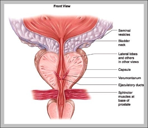
Picture of Prostate Gland. Prostate gland: A gland within the male reproductive system that is located just below the bladder. Chestnut shaped, the prostate surrounds the beginning of the urethra, the canal that empties the bladder. The prostate is actually not one but many glands, 30-50 in number, between which is abundant tissue containing…
The collagen fibers of the tissue provide strength to the tissue while the smooth muscle permits the tissue to contract to expel fluids. Fibromuscular tissue forms the outermost layer of the prostate and the tissue surrounding the urethra. Physiology of the Prostate Secretion
The fibromuscular tissue of the anterior lobe contracts to expel semen during ejaculation. The median lobeis found just posterior to the urethra along the midline of the prostate. The median lobe contains the ejaculatory ducts of the prostate. The posterior lobeforms a thin layer of tissue posterior to the median lobe and the lateral lobes.
Prostate Gland Anatomy Pictures 2