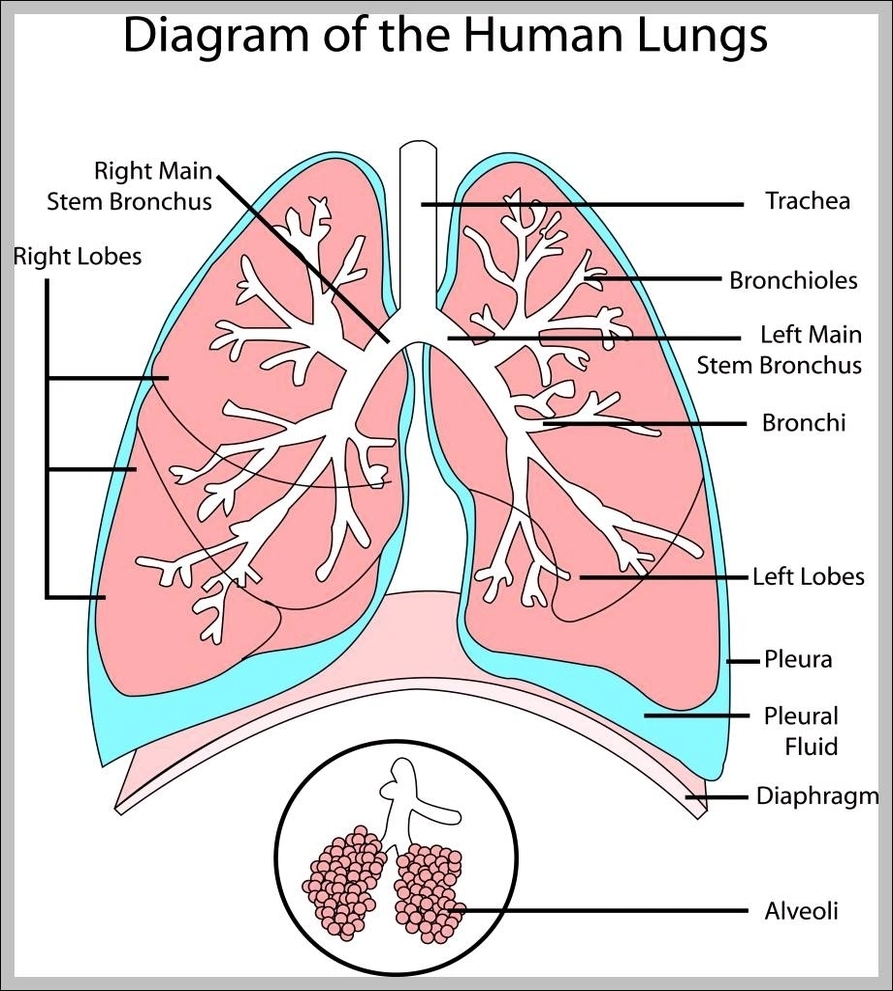
Picture of Lungs. The lungs are a pair of spongy, air-filled organs located on either side of the chest (thorax). The trachea (windpipe) conducts inhaled air into the lungs through its tubular branches, called bronchi. The bronchi then divide into smaller and smaller branches (bronchioles), finally becoming microscopic.
Picture of Lungs The lungs are a pair of spongy, air-filled organs located on either side of the chest (thorax). The trachea (windpipe) conducts inhaled air into the lungs through its tubular branches, called bronchi. The bronchi then divide into smaller and smaller branches (bronchioles), finally becoming microscopic.
The lungs are organs used for breathing located on either side of the chest. The lungs fill with air, oxygenate the blood, and dispose of carbon dioxide. Lungs are comprised of many different structures. The image on this page depicts the trachea, bronchi, and the several lobes of the left and right lungs.
Picture Of Lungs