The thoracic diaphragm, or simply the diaphragm (Ancient Greek: διάÏÏαγμα, translit. diáphragma, lit. ‘partition’), is a sheet of internal skeletal muscle in humans and other mammals that extends across the bottom of the thoracic cavity.
The diaphragm is located between the thoracic and abdominal cavities [3], with important organs like the lungs and heart located superior to it, and the liver (proximal position), kidney and stomach being inferior to it. The curved muscle is inserted into the lower part of the rib cage.
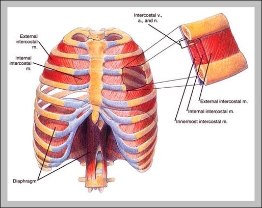
The diaphragm is shaped as two domes, with the right dome positioned slightly higher than the left because of the liver. The depression between the two domes is due to the pericardium slightly depressing the diaphragm. Thoracic surface of the diaphragm (diagram) The diaphragm has two surfaces: thoracic and abdominal.
Thoracic Diaphragm